PET/CT scans can help prevent the use of invasive biopsies in patients with suspicious growths in the mediastinum, according to a study published April 1 in the Journal of Thoracic and Cardiovascular Surgery.
A group at Stanford University in Stanford, CA, analyzed PET/CT scans to see if they could help differentiate thymomas from lymphomas, two types of cancer found in the chest cavity that are usually differentiated by biopsies. The team found PET/CT data was highly effective at identifying thymomas, which can be risky to biopsy.
"PET/CT is a reliable tool to more confidently operate without biopsy on a majority of resectable thymomas (avoiding spillage from biopsy)," wrote first author Dr. Catherine Byrd, a thoracic surgery postdoctoral fellow.
Thymoma and lymphoma are the two most common types of growths in the mediastinum, the chest cavity between the lungs. Biopsies are always required to identify lymphomas, which are treated with systemic drug therapy, while thymomas are ideally surgically removed without biopsies, which can increase the risk of spreading the cancer, according to the authors.
In this retrospective analysis, Byrd and colleagues aimed to determine whether F-18 FDG-PET/CT scans can differentiate between the thymoma and lymphoma without the need for biopsies, instead utilizing differences in maximum standardized uptake values (SUVmax) of the FDG radiotracer to distinguish between the two types.
The researchers included PET/CT imaging of 48 subjects with thymoma and 29 with anterior mediastinal lymphoma who had been treated at Stanford hospitals between 2006 and 2019. The patient group was 61% female, with a median age of 55. The median tumor size was 6.9 cm, and 21 of the 48 patients with thymomas had undergone pretreatment biopsies.
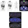
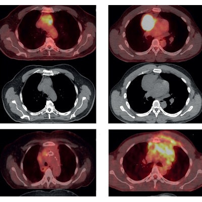
 Panel A and B demonstrate resectable disease. Panel C and D demonstrate unresectable disease that would benefit from systemic therapy. Panel A demonstrates a thymoma that appears suitable for resection on PET/CT imaging (above) and CT imaging (below). Panel B demonstrates a resectable lymphoma on PET/CT imaging (above) and CT imaging (below). Panel C demonstrates an unresectable thymoma on PET/CT imaging (above) and CT imaging (below). This thymoma is unresectable given invasion of the mass into the great vessels. Stents were placed prior to imaging to relieve compression by the mass. Panel D demonstrates an unresectable lymphoma on PET/CT imaging (above) and CT imaging (below). This lymphoma is unresectable given the direct extension of the tumor into the ribs and surrounding muscle. Image courtesy of the Journal of Thoracic and Cardiovascular Surgery.
The median SUVmax of thymoma and lymphoma differed dramatically, according to the findings: 4.35 for thymomas versus 18 for lymphomas (p < 0.001). An SUVmax < 12.85 was associated with thymoma with 100% sensitivity and approximately 89% positive predictive value, while SUVmax < 7.5 demonstrated 100% positive predictive value for thymoma.
In other words, tumors with SUVmax < 7.5 are likely thymoma and thus can be surgically removed without biopsy, while tumors with SUVmax > 7.5 should be biopsied to rule out lymphoma, the researchers suggested. Lymphoma is likely with SUVmax > 12.85, they added.
"PET/CT SUVmax of resectable anterior mediastinal masses may help guide a direct surgery vs. biopsy decision," the authors wrote.
The study was limited by a moderate sample size, which allows for the possibility that the modeling performed on the sample lacks generalizability. To address this limitation and to validate the results, a prospective multicenter study with larger patient cohort would be required, the authors wrote.
Nonetheless, PET/CT SUVmax appears to be a useful tool to help differentiate resectable thymoma from anterior mediastinal lymphoma, they suggested.
"We believe that this, in addition to the entire clinical picture, may provide a valuable guide in making the decision between direct resection and biopsy," Byrd and colleagues concluded.