An experimental PET radiotracer can visualize immune cell activity in the artery walls of patients with atherosclerosis, a finding that could lead to a new approach for early diagnosis of the disease, according to a study published March 30 in Atherosclerosis.
Danish researchers investigated whether the tracer, copper-64 (Cu-64) DOTA-AE105, could reveal the activity of macrophages involved in atherogenesis, the formation of plaque in the arteries. They found a significant uptake of the tracer in patients with arterial plaque above normal levels.
"uPAR is abundantly expressed by [mononuclear phagocyte system] cells in atherosclerotic plaques and can be visualized by the novel PET tracer Cu-64 DOTA-AE105," wrote corresponding author Dr. Andreas Kjaer, PhD, of the University of Copenhagen.
Cu-64 DOTA-AE105 is a PET radiotracer that's based on copper-64 (Cu-64) and is designed to bind to urokinase plasminogen activator receptor (uPAR) on the surface of certain immune cells, such as macrophages. These immune cells are known to be drivers of the process that leads to plaque in artery walls of atherosclerosis patients.
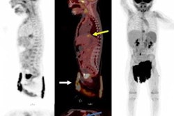
In this study, Kjaer and colleagues first used molecular biology methods to investigate uPAR expression by analyzing human atherosclerotic plaques samples and cultured cells. After the lab studies "unanimously confirmed uPAR expression" by macrophages in plaque samples, they analyzed results from whole-body PET/CT scans (Biograph mPET/CT, Siemens Healthineers) in 10 patients who underwent imaging with Cu-64 DOTA-AE105 in a previous trial.
The researchers calculated coronary artery calcium (CAC) scores for the patients and split them into low and high groups. The low CAC score group had a median score of 0 (range, 0-1) and the high CAC score group had a median score of 310 (range, 102-3871). Then, the researchers looked at uptake of the tracer in the two groups based on maximum tissue to background ratios (TBRmax) in several major arteries.
The average TBRmax value in arteries in the high CAC score group was 2.4, compared with 1.8 in the low CAC score group. A significant difference between uptake was seen in the abdominal aorta (a TBRmax of 3.2 in the high CAC group vs. 2 in the low CAC group) and in the ascending aorta (TBRmax of 2.7 in the high CAC group vs. TBRmax of 2 in the low CAC group).
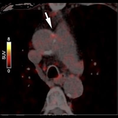
 Image courtesy of Atherosclerosis.
However, accumulation of Cu-64 DOTA-AE105 in the left and right common carotid arteries between the groups did not show a significant difference, the researchers wrote. Finally, they found higher average TBRmax values in all individual blood vessels of the high CAC score group compared to the low CAC score group.
"Heterogeneous uptake within the chosen arteries was observed on the clinical PET/CT scans. The uPAR tracer uptake was significantly higher in patients with a high coronary artery calcium score," the researchers stated.
The results of the clinical component of the study were encouraging, but the small sample size and retrospective nature were limitations of the study, the researchers wrote.
However, the preclinical component of the study was very promising, given that plaque tissue samples provide an extremely accurate and relevant platform to understand the plaque microenvironment, the researchers added.
"This retrospective analysis of clinical data combined with the in vitro and ex vivo assays show uPAR expressed by [macrophages] is involved in development of atherosclerosis that can be noninvasively assessed by Cu-64 DOTA-AE105 uPAR-PET," Kjaer and colleagues concluded.