Tuesday, December 1 | 10:40 a.m.-10:50 a.m. | SSG11-02 | Room S505AB
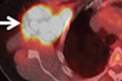
A new study from Swiss researchers is extolling the virtues of contrast-enhanced PET/MRI for patients with newly diagnosed head and neck cancer.Lead author Dr. Tetsuro Sekine, PhD, from the division of nuclear medicine at University Hospital Zurich, and colleagues concluded that contrast-enhanced PET/MRI offers diagnostic accuracy equal to that of contrast-enhanced PET/CT for this clinical application.
In the prospective study, contrast-enhanced PET/CT and PET/MRI were performed on 27 patients with newly diagnosed head and neck cancer. Tumor staging with PET/CT was correct in more than 60% of the subjects, compared with more than 70% using PET/MRI. The two hybrid modalities achieved similar sensitivity, specificity, and accuracy.
PET/MRI can be used safely for the initial workup of these patients and offers lower radiation dose than PET/CT, Sekine and colleagues concluded.
"Moreover, the MR component provides a higher soft-tissue contrast than CT, which is very helpful in the staging of local tumor extent," Sekine wrote in an email to AuntMinne.com. "Our results in a limited number of patients tell us that PET/MRI might be safely used instead of PET/CT, with a nonsignificant tendency toward better results obtained with PET/MRI."