Filtered back projection (FBP) and attenuation-weighted ordered-subsets expectation maximization (AW-OSEM) protocols have demonstrated efficacy as image reconstruction algorithms for use in 3D whole-body PET oncology imaging. A recent study from the Netherlands assessed the quantitative accuracy of each protocol in both 3D acquisition and iterative reconstruction for cardiac FDG-PET.
Researchers from the departments of nuclear medicine and PET research and cardiology at VU University Medical Centre in Amsterdam used an interleaved 2D/3D scan protocol to enable comparison of 2D and 3D imaging within the same patient and study.
In 2D PET imaging, detectors are separated by lead septa that permit coincidences to occur between opposing or adjacent detectors. In 3D PET imaging, the septa are retracted and coincidences between all possible detector banks are admitted.
The scientists, who presented their work in a poster at the 2004 Academy of Molecular Imaging conference in Orlando, FL, reconstructed the data with both FBP and AW-OSEM protocols.
"Both online continuous arterial sampling and image-derived input functions (IDIFs) were used to measure plasma curves," they wrote. "The plasma input function was determined by multiplication of the calibrated whole-blood data with a linear fit to the plasma-to-whole-blood ratios as determined from the discrete blood samples."
Online blood data was calibrated with the use of six blood samples taken during the PET scan. Eight male patients between 56 and 76 underwent a 10-minute transmission scan on an ECAT Exact HR+ PET system (Siemens Medical Solutions, Erlangen, Germany).
They were then given 370 MBq of 18F-FDG as a bolus and had a 10-minute dynamic 3D emission scan, which was followed by a series of 10 five-minute scans alternating between 2D and 3D modes, according to the authors.
 |
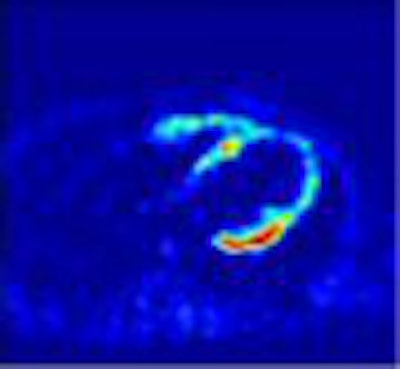
| FDG metabolic rate images derived from 2D FBP, 3D FBP, 2D OSEM, and 3D OSEM images and a plasma input function based on continuous arterial sampling. Image and caption courtesy of Mark Lubberink, Ph.D., medical physicist at the department of nuclear medicine and PET research, VU University Medical Centre. |
 |
The group used Fourier rebinning (FORE) on the 3D data (a technique that rebins 3D acquisitions into 2D sinograms, allowing it to be reconstructed with either 2D FBP or iterative methods), and reconstructed the images using FBP or AW-OSEM. The images were then subjected to a region of interest (ROI) analysis and a parametric image analysis, and IDIFs were compared to a measured online curve.
The researchers noted that the ascending aorta is the preferred choice for the definition of IDIFs, 2D, 3D, FBP, and AW-OSEM images because it resulted in metabolic rate values that had a linear correlation to those obtained with an on-line input function. As a result of their research, they determined that 3D cardiac PET acquisition is preferable to standard cardiac 2D acquisitions.
"Iterative reconstruction using AW-OSEM is a quantitatively accurate alternative to FBP for cardiac FDG-PET," they concluded. "Good correlation and no bias of metabolic rate values in relation to those based on FBP images, combined with less image noise, make 3D acquisition with FORE/AW-OSEM reconstruction a preferred choice for cardiac 18F-FDG-PET studies."
By Jonathan S. BatchelorAuntMinnie.com staff writer
July 2, 2004
Related Reading
PET/CT improves noninvasive diagnosis of cardiovascular disease, May 5, 2004
Slower CT may be better in cardiac PET/CT, March 31, 2004
Gated PET offers one-stop assessment of multiple myocardial segments, June 23, 2003
Along with SPECT, PET and MRI find niche in myocardial assessment, October 21, 2002
Copyright © 2004 AuntMinnie.com