Dutch researchers have concluded that in vivo human imaging of the right coronary artery (RCA) at 7-tesla MRI equals or surpasses 3-tesla MRI, according to a study published in the October issue of Radiology.
A comparison of the two magnet strengths in 10 healthy young adult volunteers showed that at 7 tesla the contrast-to-noise ratio between blood and epicardial fat was significantly improved, signal-to-noise ratio of the blood pool was increased, and vessel sharpness was enhanced. The clinical benefit of the improved visualization of vessel borders and greater signal-to-noise ratio is better noninvasive identification of luminal coronary artery disease without subjecting patients to ionizing radiation.
The lead author of the study, Saskia van Elderen, MD, is from the department of radiology at Leiden University Medical Center in Leiden, the Netherlands (Radiology, October 2010, Vol. 257:1, pp. 254-259).
7-tesla availability
According to the authors, 7-tesla cardiac MRI has been limited by the small number of scanners available for human use (approximately 40) and the perception that imaging at such high field strengths would be problematic due to magnetic field inhomogeneity and rules regarding specific absorption rates in humans. Also, most commercially available 7-tesla scanners do not have body radiofrequency (RF) transmit coils or surface receive coils.
However, some early studies have demonstrated that 7-tesla cardiac MRI is feasible. Van Elderen and colleagues noted, though, that they weren't aware of any studies comparing image quality at 7 tesla to that obtained at lower field strengths.
To answer that question, the researchers performed 3D MR angiography of the right coronary artery on 10 healthy young adult volunteers who had a mean age of 25 years. Seven men and three women underwent imaging at 7 tesla and 3 tesla (Achieva, Philips Healthcare, Andover, MA) in the prospective study, with 7-tesla scans performed before 3-tesla imaging in all 10 cases.
Average total scanning time was 30 minutes at 7 tesla and 20 minutes at 3 tesla. The mean interval between the two MRI scans was eight weeks. None of the subjects received nitroglycerin before MR imaging.
For the 7-tesla MR images, the researchers also used a quadrature transmit-receive surface coil, which was designed in-house and featured two overlapping loops (13-cm diameter each). On the 3-tesla MRI system, a body coil was used for RF transmission, with a commercial six-element cardiac coil array for signal reception.
Measurement parameters
To compare results from the two magnet strengths, the group measured contrast-to-noise (C/N) ratio between the blood pool and the epicardial fat, signal-to-noise (S/N) ratio of the blood pool, RCA vessel sharpness and diameter of the first 4 cm, and visible vessel length.
The researchers defined C/N ratio as the difference in signal intensity between a manually placed region of interest (ROI) in the aortic root near the offshoot of the right coronary artery, and that of an ROI placed in the epicardial fat adjacent to the proximal RCA, divided by the standard deviation of the background signal from an ROI positioned anterior to the chest wall.
Signal-to-noise ratio was calculated for the blood signal intensity in the described ROI localized in the aortic root, according to the authors. The average signal intensity from this ROI was divided by the noise.
 |
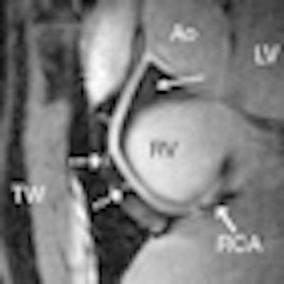
| 7-tesla (a) and 3-tesla MR (b) angiograms of the RCA in a healthy 18-year-old male show improved suppression of epicardial fat (long dotted arrow), with high contrast between the blood and epicardial fat visible at 7 tesla. At both field strengths, a number of small branching vessels are depicted (short dashed arrows). Also at 7 tesla, a long portion of the RCA is visible (solid arrow = distal part of RCA). Ao = aortic root, LV = left ventricle, RV = right ventricle, and TW = thoracic wall. All images courtesy of Radiology. |
In evaluating the MRI scans, van Elderen and colleagues found that all images displayed a high signal intensity of the coronary artery lumen, while that of the surrounding epicardial fat was suppressed. Using 7-tesla MRI, suppression of the epicardial fat was visually improved when compared with 3 tesla. In addition, the quantitative C/N ratio between the blood pool and epicardial fat was significantly improved at 7 tesla.
When compared with 3-tesla MRI, the S/N ratio of the blood pool measured on the 7-tesla images was 60% higher, with improved delineation of the right coronary artery at 7 tesla and good depiction of RCA branches and distal segments. Objective vessel sharpness analysis also demonstrated improved quantitative vessel conspicuity at 7 tesla, the authors wrote.
The group also noted the following results:
- No significant differences were found in vessel length or vessel diameter measurements among the images from the different field strengths.
- Navigator efficiency and total data acquisition time were not field-strength dependent.
- There was no significant difference in C/N ratio between the blood pool and epicardial fat, the S/N ratio of the blood pool, RCA vessel sharpness, vessel length, vessel diameter, navigator efficiency, or acquisition time between the 3-tesla images acquired with the different navigator localizations.
 |
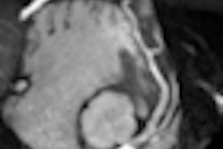
| Right coronary artery images in a healthy 26-year-old male show high visual vessel definition (dotted arrows) in the 7-tesla image (a) compared to 3 tesla (b). At 7 tesla, contrast is limited between the myocardium and the blood pool. Multiple right coronary artery side branches (dashed arrows) and distal parts of the RCA (solid arrow) are depicted clearly at both field strengths. |
Based on the images, van Elderen and colleagues concluded that quantitative parameters related to image quality attained at 7-tesla MRI equaled or surpassed those from 3-tesla in the 10 healthy young adult volunteers.
"Our results clearly warrant further evaluation in patients with coronary artery disease to assess the potential of our 7-tesla approach for the visualization of luminal RCA disease," the authors added. "Future work will concentrate on refinements in coil technology and contrast generation to support concomitant imaging of the left coronary system."
By Wayne Forrest
AuntMinnie.com staff writer
October 20, 2010
Related Reading
3-tesla 3D cardiac MRI boosts therapy planning, September 23, 2010
Cardiac MRI can help cut costs in the ER, June 29, 2010
High-field MRI appropriate for fetal autopsy, August 18, 2009
Cardiac MR techniques improve myocardial assessment, June 15, 2009
Cardiac magnetic resonance can assess at-risk myocardium after MI, June 5, 2009
Copyright © 2010 AuntMinnie.com