Abdominal aortic calcification detected on routine spine x-rays is common in women with rheumatoid arthritis, occurring in about 25% of patients, and is associated with cardiovascular disease risk. Scoring aortic calcification during routine spine imaging for vertebral fracture assessment can provide valuable information about cardiovascular and cardiometabolic burden without additional cost or radiation exposure.
- Abdominal aortic calcification (AAC) was present in 25% of 196 women with longstanding RA, with extended calcification in 13%
- AAC of any severity was associated with 2.89 times higher odds of overt cardiovascular disease
- Extended AAC was independently linked to hypertension, longer RA duration, and lower bone mineral density
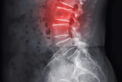
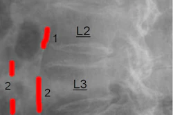
- The Kauppila score provides a validated, cost-effective method for grading AAC on lateral spine x-rays already being performed for fracture evaluation
- AAC assessment works best as a complementary indicator of cardiometabolic burden rather than a standalone screening test in RA care
Abdominal aortic calcification detected on routine spine x-rays is common among women with rheumatoid arthritis (RA) and is associated with cardiovascular disease, researchers have reported.
The finding is from a study that included 196 women with longstanding RA, and suggests that opportunistic abdominal aortic calcification (AAC) assessment may complement cardiovascular and skeletal risk evaluation in these patients.
"Kauppila-scored AAC may add clinically relevant information to conventional cardiovascular and skeletal risk assessment, particularly when spine imaging is already being performed for vertebral fracture evaluation," wrote Marcela Pinheiro, MD, of the Federal University of São Paulo in Brazil, and colleagues. The study was published November 10 in Clinical Rheumatology.
Cardiovascular disease (CVD) is a leading cause of premature mortality in RA, with patients carrying higher rates of cardiovascular morbidity and mortality than the general population, the authors explained. Traditional risk algorithms often underestimate CVD risk in patients with inflammatory joint disease, and imaging that identifies subclinical atherosclerosis is not always routinely available or appropriate, they added.
The Kauppila score is a validated method for grading AAC on lateral spine images and has been associated with cardiovascular events in the general population, but its value as an opportunistic marker in longstanding RA is unclear, the researchers noted.
To bridge the gap, the group enrolled 196 consecutive women (mean age 62.8 years old) with longstanding RA at their tertiary hospital. All participants underwent dual-energy x-ray absorptiometry scans and conventional spine x-ray. The researchers graded AAC using the Kauppila score (absent, mild-to-moderate, or extended) and used multivariable models to identify factors independently associated with AAC and overt CVD.
According to the results, AAC was present in 48 women (25%), including extended AAC in 25 (13%). Overt CVD was identified in 32 (16%). In multivariable analysis, extended AAC was independently associated with hypertension (OR, 8.57), longer RA duration (OR, 1.05 per year), and lower femoral neck T-score (OR, 0.57). AAC of any severity was associated with overt CVD (OR, 2.89).
"AAC detected on routine spine radiographs was common among women with longstanding RA and was associated with cardiovascular burden and lower femoral neck [bone mineral density]," the authors wrote.
Ultimately, from a practical standpoint, opportunistic assessment of AAC on lateral spine x-rays may be most useful as a complementary indicator of cardiometabolic burden rather than as a standalone screening test, the researchers suggested. AAC scoring could be considered when spine radiographs are already obtained for vertebral fracture assessment or other clinical indications, without substantial additional cost or radiation exposure, they wrote.
Longitudinal studies are needed to determine whether AAC progression predicts incident cardiovascular events and whether incorporating the marker into RA care pathways improves patient outcomes, the group concluded.
The full study is available here.