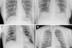
How far the crew of the recent Fram2 space mission got through the 30-page x-ray protocol they had on board is currently a mystery.
Mike Cairnie, director of global and military sales for MinXray, the developer of the ultraportable machine the astronauts used, explained that if the crew made it through even the first portion of the document, there will be a lot more to come than what we’ve seen so far.
And while the hand x-ray acquired during the mission appears to prove the feasibility of using the technology in space, as well as perhaps validates the x-ray detector’s “spectral imaging” capabilities, the focus for ultraportable x-ray now returns to Earth, Cairnie said.
“If we can put x-ray machines in space, there's really no logical reason we can't provide access, good diagnostic care, to all the people on the planet that need it,” he said.
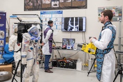
Cairnie was part of the crew in a crucial test of the x-ray machine in a zero-gravity in 2023 – on a plane colleague and consultant on the Fram2 mission Michael Pohlen, MD, recently described as a “vomit comet.”
His travels have taken him to Nepal, a number of African countries, and soon to the deepest mine in South Africa.
“We're looking at the possibility duplicating now the lowest x-ray ever taken in a mine in South Africa a couple of miles underground,” he said.
![Representative example of a 16-year-old male patient with underlying X-linked adrenoleukodystrophy. (A, B) Paired anteroposterior (AP) chest radiograph and dual-energy x-ray absorptiometry (DXA) report shows lumbar spine (L1 through L4) areal bone mineral density (BMD). The DXA report was reformatted for anonymization and improved readability. The patient had low BMD (Z score ≤ −2.0). (C) Model (chest radiography [CXR]–BMD) output shows the predicted raw BMD and Z score in comparison with the DXA reference standard, together with interpretability analyses using Shapley additive explanations (SHAP) and gradient-weighted class activation maps. The patient was classified as having low BMD, consistent with the reference standard. AM = age-matched, DEXA = dual-energy x-ray absorptiometry, RM2 = room 2, SNUH = Seoul National University Hospital, YA = young adult.](https://img.auntminnie.com/mindful/smg/workspaces/default/uploads/2026/04/ai-children-bone-density.0snnf2EJjr.jpg?auto=format%2Ccompress&fit=crop&h=100&q=70&w=100)


![Representative example of a 16-year-old male patient with underlying X-linked adrenoleukodystrophy. (A, B) Paired anteroposterior (AP) chest radiograph and dual-energy x-ray absorptiometry (DXA) report shows lumbar spine (L1 through L4) areal bone mineral density (BMD). The DXA report was reformatted for anonymization and improved readability. The patient had low BMD (Z score ≤ −2.0). (C) Model (chest radiography [CXR]–BMD) output shows the predicted raw BMD and Z score in comparison with the DXA reference standard, together with interpretability analyses using Shapley additive explanations (SHAP) and gradient-weighted class activation maps. The patient was classified as having low BMD, consistent with the reference standard. AM = age-matched, DEXA = dual-energy x-ray absorptiometry, RM2 = room 2, SNUH = Seoul National University Hospital, YA = young adult.](https://img.auntminnie.com/mindful/smg/workspaces/default/uploads/2026/04/ai-children-bone-density.0snnf2EJjr.jpg?auto=format%2Ccompress&fit=crop&h=112&q=70&w=112)


