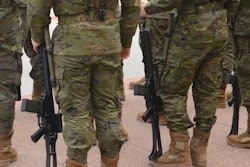
Researchers in Australia have invented a technique using x-rays and 3D surface body scans that could optimize body armor for soldiers, according to a study published May 18 in Applied Ergonomics.
The method involves using using 3D organ models based on planar x-rays and 3D external body surface scans to identify essential organ boundaries. This is fundamental to ensuring that future body armor systems provide adequate protection, noted lead author Celeste Coltman, PhD, of the University of Canberra, and colleagues.
“If this method is refined and evaluated further, it has the potential to be used as a tool for estimating body armor coverage requirements,” the group wrote.
Given that the anthropometric dimensions of soldier populations have been found to be highly variable, understanding essential organ position relative to torso anthropometry is needed to ensure that body armor provides adequate protection, the authors explained.
Yet methodologies previously adopted using CT and MRI to determine organ boundaries are limited for widespread use on large cohorts of soldiers due to their high cost and time requirements, they noted.
To develop an alternative approach, the group recruited 10 male and 10 female participants who underwent an anterior-posterior and a lateral chest/upper abdomen x-ray, 3D anthropometric surface scans of the torso, as well as MRI scans.
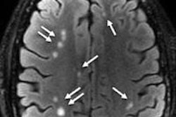
The researchers loaded the imaging data into customized Matlab software and overlaid the x-ray images on 3D organ models within each participant's surface scan to determine the position of each subject's heart, aorta, liver, and spleen. Once each organ model was positioned, a second Matlab script was used to extract data relating to organ size and position.
To evaluate the method for identifying the boundaries of essential organs, they compared it with data obtained from the MRIs.
According to the analysis, the most accurate anatomical boundaries were the left, right, and inferior boundaries of the heart, and lateral boundaries for the liver and spleen. While organ position data was significantly different between the heart model and MRI data for the left and inferior borders, the differences on average were minimal, ranging from 6.8 mm to 7.2 mm, the group noted.
Ultimately, out of the 18 participants analyzed, positioning the heart, liver, spleen, and aorta within the 3D space of each participant's surface scan was successful for 11 participants, the authors wrote.
“When the organ models were integrated within the 3D space of the participant's surface scan, visual inspection of the data suggests that this method may be suitable as a practical tool for estimating body armor coverage requirements,” the group wrote.
Ultimately, the authors suggested that x-ray and 3D surface scans could be integrated as part of basic screening undertaken for all defense personnel during recruitment and/or basic training, or as part of equipment issuing processes.
Future work will use radiopaque skin markers that appear in both the x-ray image and 3D surface scan to improve the positioning of the organ models within the 3D space as well as account for rotational error during imaging, they wrote.
“This approach represents a simpler, more cost-effective analysis and therefore, potentially, a more feasible methodology,” the investigators concluded.
The full study is available here.