Nano-X Imaging (Nanox) has received 510(k) clearance from the U.S. Food and Drug Administration (FDA) for its TAP2D cloud-enabled image enhancement for the Nanox.ARC and Nanox.ARC X digital tomosynthesis systems.
TAP2D provides radiologists with an additional 2D view generated from the digital tomosynthesis scan and automatically delivered within the existing clinical environment. The feature is designed to support evaluation while maintaining a smooth workflow for users Nanox said.
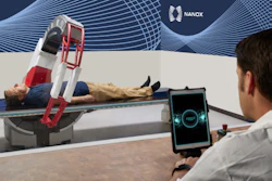
Nanox.ARC and Nanox.ARC X are FDA-cleared, multisource digital tomosynthesis systems that use 3D imaging technology to provide enhanced diagnostic capabilities at a lower cost and radiation dose than traditional systems.
Nanox said that it plans to make TAP2D available to existing installations as part of an ongoing rollout of imaging improvements.
TAP2D and other software upgrades and new capabilities can be added remotely to the Nanox.ARC systems following this and future regulatory clearances, the firm added.


![Representative example of a 16-year-old male patient with underlying X-linked adrenoleukodystrophy. (A, B) Paired anteroposterior (AP) chest radiograph and dual-energy x-ray absorptiometry (DXA) report shows lumbar spine (L1 through L4) areal bone mineral density (BMD). The DXA report was reformatted for anonymization and improved readability. The patient had low BMD (Z score ≤ −2.0). (C) Model (chest radiography [CXR]–BMD) output shows the predicted raw BMD and Z score in comparison with the DXA reference standard, together with interpretability analyses using Shapley additive explanations (SHAP) and gradient-weighted class activation maps. The patient was classified as having low BMD, consistent with the reference standard. AM = age-matched, DEXA = dual-energy x-ray absorptiometry, RM2 = room 2, SNUH = Seoul National University Hospital, YA = young adult.](https://img.auntminnie.com/mindful/smg/workspaces/default/uploads/2026/04/ai-children-bone-density.0snnf2EJjr.jpg?auto=format%2Ccompress&fit=crop&h=100&q=70&w=100)


![Representative example of a 16-year-old male patient with underlying X-linked adrenoleukodystrophy. (A, B) Paired anteroposterior (AP) chest radiograph and dual-energy x-ray absorptiometry (DXA) report shows lumbar spine (L1 through L4) areal bone mineral density (BMD). The DXA report was reformatted for anonymization and improved readability. The patient had low BMD (Z score ≤ −2.0). (C) Model (chest radiography [CXR]–BMD) output shows the predicted raw BMD and Z score in comparison with the DXA reference standard, together with interpretability analyses using Shapley additive explanations (SHAP) and gradient-weighted class activation maps. The patient was classified as having low BMD, consistent with the reference standard. AM = age-matched, DEXA = dual-energy x-ray absorptiometry, RM2 = room 2, SNUH = Seoul National University Hospital, YA = young adult.](https://img.auntminnie.com/mindful/smg/workspaces/default/uploads/2026/04/ai-children-bone-density.0snnf2EJjr.jpg?auto=format%2Ccompress&fit=crop&h=112&q=70&w=112)


