Dear AuntMinnie member,
Vendors like Siemens Healthineers have introduced camera-based and AI-powered functions in x-ray systems to support image acquisitions, yet the technology’s impact on workflows has yet to be validated in clinical routine. In an article we're featuring this week, scientists with the company say they’ve done that, and suggest use of the functions can save radiographers time. Click here for our report.
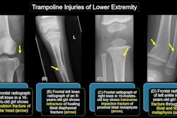
In other news, the American Roentgen Ray Society (ARRS) 2024 annual meeting was held in Boston recently and we covered several presentations for the digital x-ray community. In one, Neetika Gupta, MD, a pediatric radiologist at the University of Toronto, presented imaging evidence that suggests there may be no such thing as recreational “safe jumping” for children on trampolines.
In another story from ARRS 2024, we discussed a noncontrast fluoroscopy method developed by researchers at Vanderbilt University Medical Center in Nashville, TN, that they say can image chronic hypertension.
But AI research -- from implementing large language models in practice to developing new algorithms -- remains top in our coverage, with the following stories highlighting just some of the advances in the field:
- According to a group at New York University Langone Health, AI has delivered a long overdue update of pediatric bone growth predictions used in x-ray imaging to monitor scoliosis.
- ChatGPT-4 appears useful for extracting details from reports on thrombectomy procedures in stroke patients, according to a group in Germany.
- A team at Michigan State University developed an AI model for detecting rib fractures in children under three years old and noted that the ribcage is the most common fracture site in abused children.
- Chest dynamic digital radiography (DDR) took a step forward, with researchers developing AI to perform the time-consuming lung analysis involved in the technology.
In related news, calls are increasing for the U.S. Centers for Medicare and Medicaid Services (CMS) to sort out a payment pathway for AI applications, with less than 10 covered so far out more than 600 AI-enabled medical devices cleared by the U.S. Food and Drug Administration.
We're also featuring research that used dual energy x-ray absorptiometry scans to evaluate several patient groups. In one study, a team at Children's Hospital Colorado in Colorado Springs found that bone mineral density is only slightly below the normal average among transgender youth undergoing gender-affirming hormone therapy. In another, a group in the Netherlands found that physical activities like running may increase the risk of osteoarthritis in individuals with lower muscle mass surrounding the knee joints.
That's all for now. Be sure to check our Digital X-Ray content area often for more news.