Intelligent virtual and AI-based collimation features appear to save radiographers time during x-ray image acquisitions – a key function for enabling more patient-focused workflows, according to a recent study.
In research led by scientists at Siemens Healthineers, the use of “auto thorax collimation” for instance reduced x-ray exam lengths by 31% and the number of system interactions by 50% compared to manual collimation, noted lead author Anna Rasche, PhD, and colleagues.
“With the introduction of system and artificial intelligence functionalities in radiology, there has been a debate about their impact on the diagnostic workflow, including the radiographer's role,” the group wrote. The study was published online May 18 in Radiography.
Over the last several years, vendors like Siemens have introduced camera-based and AI-powered functions in x-ray systems to support image acquisition, with integration of such technology likely increasing in the future, the authors explained. Yet the specific impacts of the technology on the workflow in contemporary radiography rooms have not yet been validated in the clinical routine, they noted.
To that end, the researchers conducted an observational study at five clinical sites in Europe and the U.S. on the impact of features of the company’s YSIO X.pree system, a digital radiography system launched in 2020.
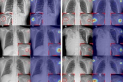
The researchers focused on potential time savings gained with the use of three features: AI-based auto thorax collimation (ATC), smart virtual ortho (SVO) collimation for stitched long-leg and full-spine exams, and virtual collimation (VC) at the radiography system workstation. Data was collected by two researchers who conducted semi-structured observations at the sites during routine exams with the features either activated or deactivated.
According to the analysis, the median exam time during 95 thorax exams was 31 seconds with ATC versus 45 seconds for 94 exams without ATC. The median number of system interactions among radiographers was one during 109 thorax examinations with ATC and two during 112 exams without ATC.
For stitched orthopedic examinations, the median exam duration was 62 seconds for 34 exams performed with SVO and 82 seconds for 40 exams without SVO, the group reported. In addition, the median time for setting the ortho range – the time between setting the upper and the lower limits of the collimation field – was 7 seconds in 39 exams with SVO versus 16 seconds in 43 exams without SVO.
Finally, VC was used to collimate in 2.4% and to check the collimation field in 68.5% of 292 observed chest and other examinations, the group noted.
“ATC and SVO enable the radiographer to save time during chest or stitched examinations. Additionally, ATC reduces machine interactions during chest examinations,” the group wrote.
Ultimately, lack of time is known to be one of the biggest stressors for radiographers during the image acquisition workflow, the authors noted. While the time savings shown per exam were relatively small in absolute numbers, the study showed that intelligent virtual and AI-based automated collimation functions can have a positive impact on the efficiency of image acquisitions, they wrote.
“In order to allow for an even more efficient image acquisition, additional workflow automation as well as the effects on the role of radiographers should be explored,” the group concluded.
The full study is available here.