
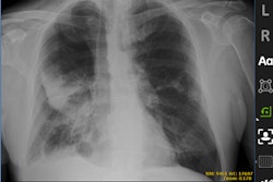
The University of California, San Francisco (USCF) said it has begun clinical use of GE Healthcare's Optima XR240amx mobile x-ray system, which features artificial intelligence (AI) algorithms developed in partnership with researchers from the university's Center for Intelligent Imaging.
The applications include an AI algorithm for pneumothorax screening that was co-developed by GE and UCSF researchers Dr. John Mongan, PhD, and Dr. Andrew Taylor, PhD. The pneumothorax algorithm received U.S. Food and Drug Administration 510(k) clearance in 2019.
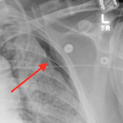
An AI algorithm developed by UCSF researchers identified a pneumothorax in a recent intensive care unit patient. The red annotations were added manually to highlight the finding. Image courtesy of Dr. John Mongan, PhD, and UCSF.Another algorithm measures the position of the endotracheal tube. It was created by Valentina Pedoia, PhD, and Sharmila Majumdar, PhD, with contributions from Dr. Thienkhai Vu, PhD, and Dr. Rutwik Shah, according to UCSF.
More algorithms developed at UCSF will be added for clinical use in the future, according to the university.


![Representative example of a 16-year-old male patient with underlying X-linked adrenoleukodystrophy. (A, B) Paired anteroposterior (AP) chest radiograph and dual-energy x-ray absorptiometry (DXA) report shows lumbar spine (L1 through L4) areal bone mineral density (BMD). The DXA report was reformatted for anonymization and improved readability. The patient had low BMD (Z score ≤ −2.0). (C) Model (chest radiography [CXR]–BMD) output shows the predicted raw BMD and Z score in comparison with the DXA reference standard, together with interpretability analyses using Shapley additive explanations (SHAP) and gradient-weighted class activation maps. The patient was classified as having low BMD, consistent with the reference standard. AM = age-matched, DEXA = dual-energy x-ray absorptiometry, RM2 = room 2, SNUH = Seoul National University Hospital, YA = young adult.](https://img.auntminnie.com/mindful/smg/workspaces/default/uploads/2026/04/ai-children-bone-density.0snnf2EJjr.jpg?auto=format%2Ccompress&fit=crop&h=100&q=70&w=100)


![Representative example of a 16-year-old male patient with underlying X-linked adrenoleukodystrophy. (A, B) Paired anteroposterior (AP) chest radiograph and dual-energy x-ray absorptiometry (DXA) report shows lumbar spine (L1 through L4) areal bone mineral density (BMD). The DXA report was reformatted for anonymization and improved readability. The patient had low BMD (Z score ≤ −2.0). (C) Model (chest radiography [CXR]–BMD) output shows the predicted raw BMD and Z score in comparison with the DXA reference standard, together with interpretability analyses using Shapley additive explanations (SHAP) and gradient-weighted class activation maps. The patient was classified as having low BMD, consistent with the reference standard. AM = age-matched, DEXA = dual-energy x-ray absorptiometry, RM2 = room 2, SNUH = Seoul National University Hospital, YA = young adult.](https://img.auntminnie.com/mindful/smg/workspaces/default/uploads/2026/04/ai-children-bone-density.0snnf2EJjr.jpg?auto=format%2Ccompress&fit=crop&h=112&q=70&w=112)


