
Researchers at the University Hospitals Cleveland Medical Centers said they have completed testing of GE Healthcare's Critical Care Suite artificial intelligence (AI) software on the company's Optima XR240amx mobile x-ray system.
GE selected the medical center in November 2019 as the first hospital in the U.S. to study Optima XR240amx with Critical Care Suite, an AI-based application designed to identify urgent cases such as pneumothorax and flag these exams for urgent review, according to the institution. The Cleveland researchers completed the evaluation phase of Critical Care Suite in December, noting the technology flagged seven to 15 collapsed lungs per day.
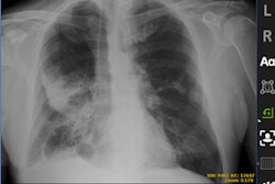
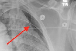
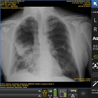
A demonstration of Critical Care Suite. Image courtesy of University Hospitals.The system has now been placed into daily clinical use, and principal investigator Dr. Amit Gupta said in a press release that the technology has helped improve workflow and patient care.
The group is now using the system to aid with COVID-19 imaging. It travels to different intensive care units (ICU) at the medical center, and future research with GE could improve ICU care for patients with COVID-19, according to University Hospitals.


![Representative example of a 16-year-old male patient with underlying X-linked adrenoleukodystrophy. (A, B) Paired anteroposterior (AP) chest radiograph and dual-energy x-ray absorptiometry (DXA) report shows lumbar spine (L1 through L4) areal bone mineral density (BMD). The DXA report was reformatted for anonymization and improved readability. The patient had low BMD (Z score ≤ −2.0). (C) Model (chest radiography [CXR]–BMD) output shows the predicted raw BMD and Z score in comparison with the DXA reference standard, together with interpretability analyses using Shapley additive explanations (SHAP) and gradient-weighted class activation maps. The patient was classified as having low BMD, consistent with the reference standard. AM = age-matched, DEXA = dual-energy x-ray absorptiometry, RM2 = room 2, SNUH = Seoul National University Hospital, YA = young adult.](https://img.auntminnie.com/mindful/smg/workspaces/default/uploads/2026/04/ai-children-bone-density.0snnf2EJjr.jpg?auto=format%2Ccompress&fit=crop&h=100&q=70&w=100)


![Representative example of a 16-year-old male patient with underlying X-linked adrenoleukodystrophy. (A, B) Paired anteroposterior (AP) chest radiograph and dual-energy x-ray absorptiometry (DXA) report shows lumbar spine (L1 through L4) areal bone mineral density (BMD). The DXA report was reformatted for anonymization and improved readability. The patient had low BMD (Z score ≤ −2.0). (C) Model (chest radiography [CXR]–BMD) output shows the predicted raw BMD and Z score in comparison with the DXA reference standard, together with interpretability analyses using Shapley additive explanations (SHAP) and gradient-weighted class activation maps. The patient was classified as having low BMD, consistent with the reference standard. AM = age-matched, DEXA = dual-energy x-ray absorptiometry, RM2 = room 2, SNUH = Seoul National University Hospital, YA = young adult.](https://img.auntminnie.com/mindful/smg/workspaces/default/uploads/2026/04/ai-children-bone-density.0snnf2EJjr.jpg?auto=format%2Ccompress&fit=crop&h=112&q=70&w=112)


