
Carestream Health has unveiled DRX-L, a new digital radiography (DR) detector designed for long-length imaging applications.
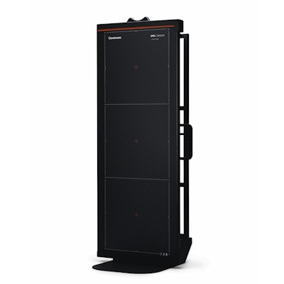
Carestream Health's new DRX-L digital radiography detector for long-length imaging. Image courtesy of Carestream Health.Now available in the U.S. and Canada, DRX-L enables hospitals and imaging centers to capture long-length images with a single exposure, providing a large field of view and high resolution for leg-length and spine exams, according to the vendor. By acquiring the image in one exposure, the detector decreases radiation dose and overall exam time compared with studies that require multiple exposures, Carestream said.
DRX-L also features the firm's ImageView software with the Eclipse image processing application. What's more, it includes Carestream's X‑Factor technology, enabling its use with other compatible DRX equipment, according to the company.
It can be utilized with Carestream digital x‑ray products such as the DRX‑Evolution Plus/DRX‑Evolution, DRX‑Ascend, Q-Rad X‑ray, and DRX‑1 digital x‑ray systems.
![Representative example of a 16-year-old male patient with underlying X-linked adrenoleukodystrophy. (A, B) Paired anteroposterior (AP) chest radiograph and dual-energy x-ray absorptiometry (DXA) report shows lumbar spine (L1 through L4) areal bone mineral density (BMD). The DXA report was reformatted for anonymization and improved readability. The patient had low BMD (Z score ≤ −2.0). (C) Model (chest radiography [CXR]–BMD) output shows the predicted raw BMD and Z score in comparison with the DXA reference standard, together with interpretability analyses using Shapley additive explanations (SHAP) and gradient-weighted class activation maps. The patient was classified as having low BMD, consistent with the reference standard. AM = age-matched, DEXA = dual-energy x-ray absorptiometry, RM2 = room 2, SNUH = Seoul National University Hospital, YA = young adult.](https://img.auntminnie.com/mindful/smg/workspaces/default/uploads/2026/04/ai-children-bone-density.0snnf2EJjr.jpg?auto=format%2Ccompress&fit=crop&h=100&q=70&w=100)


![Representative example of a 16-year-old male patient with underlying X-linked adrenoleukodystrophy. (A, B) Paired anteroposterior (AP) chest radiograph and dual-energy x-ray absorptiometry (DXA) report shows lumbar spine (L1 through L4) areal bone mineral density (BMD). The DXA report was reformatted for anonymization and improved readability. The patient had low BMD (Z score ≤ −2.0). (C) Model (chest radiography [CXR]–BMD) output shows the predicted raw BMD and Z score in comparison with the DXA reference standard, together with interpretability analyses using Shapley additive explanations (SHAP) and gradient-weighted class activation maps. The patient was classified as having low BMD, consistent with the reference standard. AM = age-matched, DEXA = dual-energy x-ray absorptiometry, RM2 = room 2, SNUH = Seoul National University Hospital, YA = young adult.](https://img.auntminnie.com/mindful/smg/workspaces/default/uploads/2026/04/ai-children-bone-density.0snnf2EJjr.jpg?auto=format%2Ccompress&fit=crop&h=112&q=70&w=112)


