
Carestream Health has introduced Lux 35, the firm's first glass-free cesium digital radiography (DR) detector for medical imaging applications.
Building off the success of Carestream's glass-free detector in the nondestructive testing market, Lux 35 is a 14 x 17-inch (35 cm x 43-cm) wireless detector that weighs around 5 lb and can be used by radiographers to perform bedside exams, according to the vendor. Lux 35 is the company's lightest detector to date.
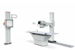
Carestream's new Lux 35 glass-free cesium detector. Image courtesy of Carestream.The detector employs the company's ImageView software powered by Eclipse and also features Carestream's X-Factor technology to support sharing with other Carestream DRX equipment, the vendor said. In addition, it supports image-processing options such as tube and line visualization, pneumothorax visualization, EVP Plus software, bone suppression, and more, according to Carestream.
The detector's LED lights and graphical user interface provide radiographers with information on its status. The firm also noted that the Lux 35's battery is backward compatible; it works with Carestream's DRX Plus detectors and utilizes the same battery charger.
Carestream is also highlighting the cesium detector's image quality and lower radiation dose, as well as its ergonomic benefits.
![Representative example of a 16-year-old male patient with underlying X-linked adrenoleukodystrophy. (A, B) Paired anteroposterior (AP) chest radiograph and dual-energy x-ray absorptiometry (DXA) report shows lumbar spine (L1 through L4) areal bone mineral density (BMD). The DXA report was reformatted for anonymization and improved readability. The patient had low BMD (Z score ≤ −2.0). (C) Model (chest radiography [CXR]–BMD) output shows the predicted raw BMD and Z score in comparison with the DXA reference standard, together with interpretability analyses using Shapley additive explanations (SHAP) and gradient-weighted class activation maps. The patient was classified as having low BMD, consistent with the reference standard. AM = age-matched, DEXA = dual-energy x-ray absorptiometry, RM2 = room 2, SNUH = Seoul National University Hospital, YA = young adult.](https://img.auntminnie.com/mindful/smg/workspaces/default/uploads/2026/04/ai-children-bone-density.0snnf2EJjr.jpg?auto=format%2Ccompress&fit=crop&h=100&q=70&w=100)


![Representative example of a 16-year-old male patient with underlying X-linked adrenoleukodystrophy. (A, B) Paired anteroposterior (AP) chest radiograph and dual-energy x-ray absorptiometry (DXA) report shows lumbar spine (L1 through L4) areal bone mineral density (BMD). The DXA report was reformatted for anonymization and improved readability. The patient had low BMD (Z score ≤ −2.0). (C) Model (chest radiography [CXR]–BMD) output shows the predicted raw BMD and Z score in comparison with the DXA reference standard, together with interpretability analyses using Shapley additive explanations (SHAP) and gradient-weighted class activation maps. The patient was classified as having low BMD, consistent with the reference standard. AM = age-matched, DEXA = dual-energy x-ray absorptiometry, RM2 = room 2, SNUH = Seoul National University Hospital, YA = young adult.](https://img.auntminnie.com/mindful/smg/workspaces/default/uploads/2026/04/ai-children-bone-density.0snnf2EJjr.jpg?auto=format%2Ccompress&fit=crop&h=112&q=70&w=112)


