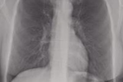
In a face-off of two digital chest x-ray techniques, dual-energy imaging proved better than a bone-suppression algorithm for detecting small lung cancers on computed radiography (CR) images. But the difference was small, and bone suppression may still have some advantages, according to an article published September 22 in Radiology.
Both dual-energy imaging and bone suppression reconstruction offer more high-tech ways to process digital chest images than conventional radiography, a technology more than 100 years old. Dual-energy uses images acquired at different energy levels to highlight either bone or soft tissue. Meanwhile, bone suppression uses software algorithms to process image data and automatically remove bone structures that might obscure pathology.
Pioneering work on both techniques has been conducted at the University of Chicago, and in the Radiology study a research group led by Dr. Feng Li, PhD, and Dr. Heber MacMahon decided to compare the two techniques to see if bone suppression might offer an alternative to dual-energy imaging (Radiology, September 22). The latter technology does have several drawbacks, including a slightly higher radiation dose and the need to use special equipment.
To create a patient population for their study, the researchers combined two separate databases; the first consisted of a group of 94 patients with 105 peripheral nodular cancers who were imaged at the university from January 2004 to September 2008. Images included both standard and dual-energy radiographs, and cancers were confirmed on CT. The second database consisted of 19 patients with 22 peripheral nodular cancers, and also included conventional and standard dual-energy images.
The researchers decided to exclude patients with obvious nodules, as well as those with poor image quality or underlying abnormalities, leaving them with a population of 50 patients with 55 nodular cancers visible on dual-energy images. They added to the population 30 patients without cancer. All radiographs were acquired with a single-exposure CR unit (Fujifilm Medical Systems USA) using 110 kVp and 2.5-16 mAs settings.
Bone suppression imaging was performed by applying a commercially available software algorithm (SoftView version 2.0, Riverain Medical) to all 80 chest images. The algorithm uses image normalization, feature extraction, and regression-network techniques to predict the bone image and then subtract that data to form the soft-tissue image, the authors wrote.
To assess the diagnostic performance of radiologists using either technique, Li and colleagues tried to replicate the workflow that might be used in a typical practice using advanced digital x-ray visualization (although the researchers noted that most practices would not use both bone suppression and dual energy simultaneously in the same exam). A group of 10 radiologists first reviewed the conventional posterior-anterior (PA) radiographs without image processing, then conventional and bone suppression images together, and then standard and dual-energy images.
 |
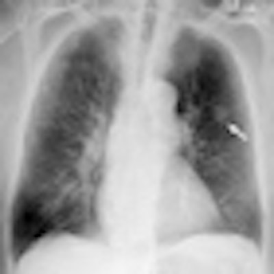
| Chest PA radiographs in 79-year-old man with nodular cancer in left upper lobe that was overlooked at the time of initial interpretation. Image at left is standard x-ray that shows cancer (arrow) partly overlapped with ribs. Bone suppression image at right shows the cancer but also enhances other structures, especially in the apical areas. Images courtesy of RSNA. |
Readers were asked to note for each image their confidence level that it included the presence of cancer. The researchers analyzed the accuracy of the readers' confidence levels against the actual presence of cancer using receiver operator characteristics (ROC) methodology, rendered as values in the area under the ROC curve (AUC). Findings in the primary study are below.
Accuracy of x-ray visualization techniques
|
The researchers wrote that the differences were statistically significant between x-ray alone and x-ray plus bone suppression (p < 0.001), between standard x-ray alone and x-ray plus dual energy (p < 0.001), and between x-ray plus bone suppression and x-ray plus dual energy (p < 0.001).
The advantage found with dual-energy over bone suppression could be accounted for in part by the fact that the study's readers were using the bone suppression software for the first time during the trial, according to Li and colleagues. The current version of the bone suppression software also increases the prominence of normal vascular structures and small scars.
The researchers concluded by stating that bone suppression is not intended to replace standard x-ray imaging for applications outside of detecting chest nodules, nor would it replace dual-energy imaging in cases where the technique is currently being used. However, they believe it could have value for the large majority of digital x-ray cases acquired without energy subtraction.
"While [dual-energy] radiography provides the highest accuracy, use of [bone suppression] software in combination with a standard radiograph can significantly improve detection of small nodular cancers compared with use of standard radiographs alone," they wrote.
| Funding for the study was provided by Riverain Medical. Dr. MacMahon is a consultant to the company. |