Radiography vendor Shimadzu of Torrance, CA, will feature a new line of flat-panel digital cardiac angiography systems, as well as the company's radiography/fluoroscopy (R/F) offerings.
BRANSIST Safire is a new digital cardiac angiography system using a 9-inch direct conversion digital flat-panel detector. The system is available in both single and biplane configurations, and a 17-inch vascular system is scheduled to be added to the product line in 2007.
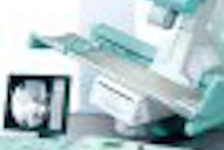
Also look for Shimadzu to highlight its Sonialvision Versa R/F table. The system is a universal positioner that covers applications from gastrointestinal exams to limited angiographic studies, and includes variable table height for patient loading and unloading.
Sonialvision Versa is available with a 12- or 16-inch image intensifier, or Shimadzu's Safire flat-panel digital detector. The system can be configured for bariatric studies with an optional patient table capable of support patients weighing up to 700 lb.
Shimadzu Medical Systems' conventional table R/F system has been redesigned as the Fluorospeed 300. This new system integrates a new generator with a large LCD console and an intelligent overhead tube mount, also with a large LCD display.
By Robert Bruce
AuntMinnie.com contributing writer
October 30, 2006
Copyright © 2006 AuntMinnie.com


![Representative example of a 16-year-old male patient with underlying X-linked adrenoleukodystrophy. (A, B) Paired anteroposterior (AP) chest radiograph and dual-energy x-ray absorptiometry (DXA) report shows lumbar spine (L1 through L4) areal bone mineral density (BMD). The DXA report was reformatted for anonymization and improved readability. The patient had low BMD (Z score ≤ −2.0). (C) Model (chest radiography [CXR]–BMD) output shows the predicted raw BMD and Z score in comparison with the DXA reference standard, together with interpretability analyses using Shapley additive explanations (SHAP) and gradient-weighted class activation maps. The patient was classified as having low BMD, consistent with the reference standard. AM = age-matched, DEXA = dual-energy x-ray absorptiometry, RM2 = room 2, SNUH = Seoul National University Hospital, YA = young adult.](https://img.auntminnie.com/mindful/smg/workspaces/default/uploads/2026/04/ai-children-bone-density.0snnf2EJjr.jpg?auto=format%2Ccompress&fit=crop&h=100&q=70&w=100)


![Representative example of a 16-year-old male patient with underlying X-linked adrenoleukodystrophy. (A, B) Paired anteroposterior (AP) chest radiograph and dual-energy x-ray absorptiometry (DXA) report shows lumbar spine (L1 through L4) areal bone mineral density (BMD). The DXA report was reformatted for anonymization and improved readability. The patient had low BMD (Z score ≤ −2.0). (C) Model (chest radiography [CXR]–BMD) output shows the predicted raw BMD and Z score in comparison with the DXA reference standard, together with interpretability analyses using Shapley additive explanations (SHAP) and gradient-weighted class activation maps. The patient was classified as having low BMD, consistent with the reference standard. AM = age-matched, DEXA = dual-energy x-ray absorptiometry, RM2 = room 2, SNUH = Seoul National University Hospital, YA = young adult.](https://img.auntminnie.com/mindful/smg/workspaces/default/uploads/2026/04/ai-children-bone-density.0snnf2EJjr.jpg?auto=format%2Ccompress&fit=crop&h=112&q=70&w=112)


