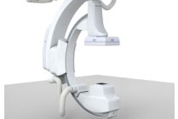
Lodox Systems of South Lyon, MI, will demonstrate Rad-MPTM and Lucid, functional enhancements to its Statscan DR system, which has been installed in 18 facilities worldwide in the past year.
The Rad-MPTM upgrade supports increased functionality in trauma and ER x-ray settings. It consists of a fully adjustable, flexible 20 x 28-inch (50 x 71-cm) radiolucent imaging table that can be used in both horizontal and vertical planes. It also includes a hydraulic patient imaging chair and pedestal stand for imaging patients in upright sitting and standing positions. This optional upgrade is priced at $25,000.
Lucid is a new image enhancement tool for Statscan that improves visualization of the lateral cervical-thoracic spine junction by nearly 100% compared to conventional 2D radiography, according to Lodox.
Lucid has FDA clearance and is available in the U.S. and worldwide. It will be a standard feature on Statscan at no additional cost.
By Robert Bruce
AuntMinnie.com contributing writer
November 8, 2005
Copyright © 2005 AuntMinnie.com


![Representative example of a 16-year-old male patient with underlying X-linked adrenoleukodystrophy. (A, B) Paired anteroposterior (AP) chest radiograph and dual-energy x-ray absorptiometry (DXA) report shows lumbar spine (L1 through L4) areal bone mineral density (BMD). The DXA report was reformatted for anonymization and improved readability. The patient had low BMD (Z score ≤ −2.0). (C) Model (chest radiography [CXR]–BMD) output shows the predicted raw BMD and Z score in comparison with the DXA reference standard, together with interpretability analyses using Shapley additive explanations (SHAP) and gradient-weighted class activation maps. The patient was classified as having low BMD, consistent with the reference standard. AM = age-matched, DEXA = dual-energy x-ray absorptiometry, RM2 = room 2, SNUH = Seoul National University Hospital, YA = young adult.](https://img.auntminnie.com/mindful/smg/workspaces/default/uploads/2026/04/ai-children-bone-density.0snnf2EJjr.jpg?auto=format%2Ccompress&fit=crop&h=100&q=70&w=100)


![Representative example of a 16-year-old male patient with underlying X-linked adrenoleukodystrophy. (A, B) Paired anteroposterior (AP) chest radiograph and dual-energy x-ray absorptiometry (DXA) report shows lumbar spine (L1 through L4) areal bone mineral density (BMD). The DXA report was reformatted for anonymization and improved readability. The patient had low BMD (Z score ≤ −2.0). (C) Model (chest radiography [CXR]–BMD) output shows the predicted raw BMD and Z score in comparison with the DXA reference standard, together with interpretability analyses using Shapley additive explanations (SHAP) and gradient-weighted class activation maps. The patient was classified as having low BMD, consistent with the reference standard. AM = age-matched, DEXA = dual-energy x-ray absorptiometry, RM2 = room 2, SNUH = Seoul National University Hospital, YA = young adult.](https://img.auntminnie.com/mindful/smg/workspaces/default/uploads/2026/04/ai-children-bone-density.0snnf2EJjr.jpg?auto=format%2Ccompress&fit=crop&h=112&q=70&w=112)


