A "quadruple low" photon-counting CT (PCCT) protocol for lung cancer imaging improves image quality and reduces radiation dose exposure to the kidneys compared with conventional CT, according to a study published February 17 in Radiology.
The findings could translate to better care for patients who require repeated contrast-enhanced imaging, wrote a team led by Xiaofei Yang, MD, PhD, of the First Affiliated Hospital of Zhengzhou University in China.
"[We found that] compared with [traditional] CT, PCCT with a quadruple-low protocol reduced the incidence of contrast-induced nephropathy and enhanced image quality and diagnostic confidence in imaging features in patients with lung cancer," the group noted.
CT plays a critical role in lung cancer diagnosis and treatment monitoring, but traditional CT has well-known limitations, including lower spatial resolution and higher radiation dose; PCCT has been shown to overcome these limitations, the researchers wrote.
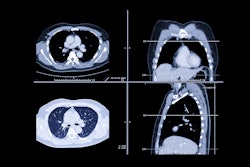
In a study that included 425 patients with lung cancer, the group compared the diagnostic quality of chest CT images from PCCT using a quadruple-low protocol with that of traditional CT imaging. Patients underwent either exam between July and September 2024, and the PCCT protocol consisted of a 2 mL/sec injection rate and 1 mL/kg of 320 mg of iodine per milliliter. Two radiologists evaluated the exams' image quality and lesion imaging features, and assessed lesion and parenchymal metrics such as attenuation, signal-to-noise ratio, and contrast-to-noise ratio.
The researchers reported the following:
- PCCT reduced radiation dose by 55% with (3.49 mSv vs. 7.82 mSv; p < 0.001).
- It reduced contrast load, with a 33% lower injection rate, 22% lower volume, and 9% lower concentration.
- No cases of contrast‑induced nephropathy occurred in the PCCT group, compared with 7.9% in the traditional CT group (p = 0.02).
- PCCT showed higher signal-to-noise ratio and contrast-to-noise ratio for lung lesions and parenchyma across noncontrast, arterial, and venous phases (all, p < 0.001).
- PCCT demonstrated superior subjective image quality, including sharpness, artifact reduction, and noise suppression (all p < 0.001).
- PCCT elicited greater diagnostic confidence from the readers for key malignant features such as lobulation, spiculation, pleural retraction, vascular convergence, and vacuoles.
 Representative imaging features of the lesion with energy-integrating detector (EID) CT and photon-counting CT (PCCT). (A) Unenhanced PCCT image (left column) in a 56-year-old man with lung cancer and (B) EID CT image (right column) in a 73-year-old man with lung cancer depict lobulation. (C) Unenhanced PCCT image (left column) in a 67-year-old man with lung cancer and (D) EID CT image (right column) in a 71-year-old woman with lung cancer depict spiculation. (E) Contrast-enhanced PCCT image (left column) in a 63-year-old man with lung cancer and (F) EID CT image (right column) in a 69-year-old woman with lung cancer depict vessel convergence. (G) Unenhanced PCCT image (left column) in a 64-year-old man and (H) EID CT image (right column) in a 49-year-old man with lung cancer depict vacuoles. (I) Unenhanced PCCT image (left column) in a 62-year-old man and (J) EID CT image (right column) in a 72-year-old man with lung cancer depict pleural retraction. The lung cancer lesions and their specific imaging features are shown in the orange boxes. In brief, compared with EID CT (B, D), PCCT provides superior visualization of contour bulge (A) and thick strands extending from the lesion edge (C), thereby enhancing the diagnostic confidence in identifying lobulation and spiculation. Vessel convergence and vacuoles are clearly depicted on PCCT images (E, G), but they appear blurred or indiscernible on EID CT images (F, H). Additionally, PCCT (I) shows clearer linear attenuation pulling of the fissure toward the lesion compared with EID CT (J), potentially improving the diagnostic confidence in pleural retraction.RSNA
Another benefit of PCCT for this indication is that improved lesion conspicuity and higher diagnostic confidence may support more accurate staging and characterization of invasive tumor features, which can influence therapeutic decisions such as surgical planning or neoadjuvant therapy selection, according to the authors.
"The combination of PCCT and the quadruple-low scanning protocol may provide a safer and more effective imaging strategy for lung cancer," they concluded.
Access the full study here.