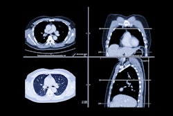
Chest CT performed for other indications can help predict hip fracture risk by measuring pectoral muscle index (PMI) and vertebral body attenuation values, researchers have reported.
Using CT in this way shows an area under the receiver operating curve of 0.86, wrote a team led by Xiong Yi Wang, MD, of the Second Affiliated Hospital of Soochow University in Suzhou, China. The study results were published September 3 in Orthopaedic Surgery.
"[We found that] compared with the healthy control group, the hip fracture group was older and had lower [body mass index], [bone mineral density], and T4HU [fourth thoracic vertebra], and [pectoral muscle index volume]," the team explained.
Hip fractures can be devastating for elderly people, and assessing risk is key to keeping this population healthy, the group wrote.
"Fragility fractures of the hip, also known as low-energy fractures, have high morbidity, mortality, and disability rates," the team noted. "It is also referred to as 'the last fracture in life.' "
Dual-energy x-ray absorptiometry (DEXA) is considered the gold standard for assessing hip fracture risk, but it can be expensive, which reduces its accessibility. In fact, more than 80% of patients with hip fracture do not undergo DEXA to assess their risk, the team explained. That's where opportunistic chest CT could come in.
"Studies have demonstrated that the … CT value of vertebrae is closely related to bone mineral density and fracture," it wrote. "This means that the CT value can be used to assess the bone mass of patients and predict fracture risk."
Wang's team sought to clarify whether chest CT imaging data -- specifically images from the fourth thoracic vertebra (T4HU) -- could opportunistically help predict patients' hip fracture risk in patients via a study that included 472 individuals who had both bone mineral density testing and chest CT between January 2021 and January 2024. Study participants were divided into a healthy control group and a hip fracture group. The researchers developed a predictive model based on the CT and PMI findings and assessed its efficacy using the receiver operating characteristic (ROC) curve.
The investigators found the following:
- Both PMI and T4HU volumes were lower in the hip fracture group than in the healthy control group (p < 0.05).
- Low PMI and low T4HU were risk factors for hip fracture.
- The predictive model that blended both PMI and T4HU data on the basis of age and BMI had an area under the curve (AUC) of 0.865 (p < 0.01) for diagnostic efficacy. It had a sensitivity of 82% and a specificity of 75%.
The study results are good news for patients at risk of hip fracture, according to the authors.
"[Our work] demonstrates for the first time that pre-existing chest CT can be used for opportunistic screening for hip fracture risk in the absence of bone mineral density [information], thereby reducing healthcare costs, rationalizing the allocation of healthcare resources, and obtaining a better cost-benefit ratio," they concluded.
The complete research can be found here.


