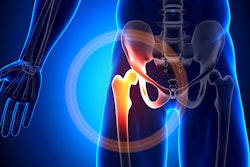
A deep-learning model that includes digital x-rays reconstructed from 3D hip CT images shows promise for predicting subsequent fractures in patients with a relatively recent hip break, researchers have found.
The findings could improve how clinicians care for their patients, wrote a team led by doctoral candidate Yisak Kim of Seoul National University Graduate School in South Korea. The group's findings were published January 30 in Radiology.
"Developing a prediction model for short-term subsequent fracture risk is important because it would identify patients at the highest risk and aid in determining appropriate treatment strategies," the team noted.
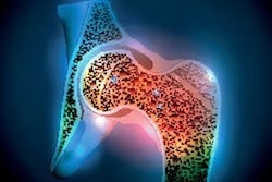
Patients who sustain an initial fracture are at highest risk for another over the next few years, the investigators explained. But models to predict short-term subsequent risk have not been developed. To fill this knowledge gap, Kim and colleagues developed and validated a deep-learning prediction model for subsequent fracture risk using digitally reconstructed x-rays from hip CT images in patients with recent hip fractures.
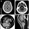
Their research included 1,480 adults who underwent 3D hip CT imaging prompted by a fracture between January 2004 and December 2020. Mean follow-up time was 3.4 years, and primary outcomes were subsequent bone fractures in the spine, hip, humerus, or wrist, occurring at least three months after the initial break. The team generated 2D frontal, lateral, and axial digitally reconstructed x-rays from the CT images, and used DenseNet convolutional neural network modules to estimate a patient's future fracture risk based on imaging features (this combination was dubbed the "ensemble model").
The researchers assessed the model's performance using the C (concordance) index (reference, 1), and the area under the receiver operating curve (AUC), comparing it with three other models. (Model A included age and sex; model B included age, sex, body mass index, smoking and alcohol consumption status, steroid use, presence of rheumatoid arthritis, and secondary osteoporosis; and model C included all of these, plus hip fracture type, femoral neck bone mineral density, and use of antiosteoporosis medication.)
Of the total patient cohort, 1,012 made up a training and validation set (and included 113 subsequent fractures); 468 made up a test set (22 subsequent fractures).
The team found the following:
| Ensemble model performance for predicting subsequent fractures, test set, compared with three other models | ||
|---|---|---|
| Measure | Other imaging models | Ensemble model |
| C index (with 1 as reference) | ||
| Overall | 0.61 to 0.65 | 0.73 |
| AUC | ||
| 2-year follow-up | 0.58 to 0.65 | 0.74 |
| 3-year follow-up | 0.64 to 0.68 | 0.74 |
| 5-year follow-up | 0.66 to 0.71 | 0.77 |
"In patients with recent hip fractures, the ensemble deep-learning model using digital reconstructed radiographs from hip CT showed good performance for predicting subsequent fractures in the short term," they concluded.
Future research is needed, but the study findings show promise for tailoring patient care, wrote Matthew Li, MD, and Jacob Jaremko, MD, PhD, both of the University of Alberta Hospital in Edmonton, Canada, in an accompanying editorial.
"While there are concerns regarding the level of performance and interpretability of the deep learning CT–based model for subsequent fracture prediction reported by … Kim et al, their results are a meaningful step toward personalizing short-term fracture prevention after hip fracture," they wrote.
The complete study can be found here.


![A normal mammogram confirmed by three-year radiologic follow-up illustrates reader-marked regions of interest (ROIs) during (A) unaided (round 1) and (B) artificial intelligence (AI)–assisted (round 2) reading. Each colored dot represents an ROI for recall by a human reader. Readers could mark more than one ROI per case, represented by multiple dots of the same color. During AI-assisted reading, the AI system displayed three visible prompts: two with suspicion of malignancy scores of 35% (left mediolateral oblique [L MLO] and craniocaudal [L CC]) and one with a suspicion of malignancy score of 10% (right craniocaudal [R CC]), shown as polygonal overlays. Without AI, six of 10 readers (60%) marked a false-positive ROI. With AI assistance, this fell to two of 10 (20%). R MLO = right mediolateral oblique.](https://img.auntminnie.com/mindful/smg/workspaces/default/uploads/2026/07/2026-07-14-radiology-mammogram-ai-auto-bias.H0bYO8QlWs.jpg?auto=format%2Ccompress&dpr=2&fit=crop&h=167&q=70&w=250)


