Photon-counting CT (PCCT) produces diagnostic CT pulmonary angiography (CTPA) images of pulmonary embolism (PE) at a low contrast dose, a study published June 29 in Academic Radiology has found.
A group led by Saher Saeed of Ruhr University Bochum in Germany found that the reduction is almost half of the highest dose administered in the study -- 35 ml compared with 60 ml.
"Significant contrast media dose reduction is possible [with PCCT] without a reduction in image quality," the team noted.
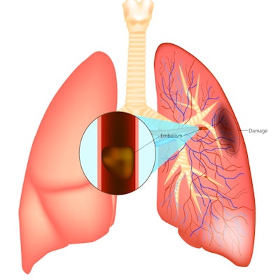
Since the 1990s, CTPA has been the go-to exam for identifying PE, a condition that can be deadly, the group explained; prognosis depends on timely start of treatment. Yet CT contrast can lead to adverse reactions such as flushed, itchy, welted, or swollen skin; low blood pressure; loss of consciousness; or even bronchospasm.
Additionally, clinicians have long been concerned about the potential of iodinized contrast to injure the kidneys. Reducing contrast dose may be key to avoiding adverse reactions, and photon-counting CT shows promise for doing this, the investigators wrote. But does reduced dose translate to reduce image quality?
To explore this question, Saeed and colleagues conducted a study that included 105 patients who underwent CTPA using contrast (Accupaque 300, GE HealthCare) and high-pitch dual-source scanning (fast low-angle shot, or FLASH mode) on a PCCT device (Naeotom Alpha, Siemens Healthineers). Patients were divided into the following groups:
- Group 1 (29): 35 ml of contrast
- Group 2 (62): 45 ml of contrast
- Group 3 (14): 60 ml of contrast
Four readers assessed image quality using the five-point Likert scale (with 1 equal to poor and 5 equal to excellent). There were no complications among study participants related to the CT exam itself or the contrast.
The team found that the lowest contrast group had the highest image quality rating, as well as statistical significance between Groups 1 and 3 (p < 0.001) and between Groups 2 and 3 (p = 0.003).
| Image quality by contrast group on a five-point scale | |||
| Measure | Group 1 (35 ml of contrast) | Group 2 (45 ml of contrast) | Group 3 (60 ml of contrast) |
| Image quality | 4.6 | 4.5 | 4.1 |
The authors also reported that most pulmonary arteries could be assessed adequately without significant differences among the groups.
The study findings are good news, as reduced contrast dose is better for patients, according to the researchers.
"PCCT CTPA using a high-pitch protocol allows to acquire excellent diagnostic imaging of the pulmonary arteries with only 35 ml [of] contrast media," they concluded.
Click here for the complete study.