Photon-counting CT (PCCT) pulmonary angiography imaging outperforms conventional CT for diagnosing pulmonary embolism (PE) -- cutting radiation dose in half, according to research presented at the RSNA meeting in Chicago.
"Photon-counting detector CT offers the possibility to gain spectral information from high-pitch CT pulmonary angiography scans which is particularly beneficial in patients with suspected PE, who frequently present with dyspnea," Pauline Pannenbecker of Uniklinikum Würzburg in Germany told session attendees.
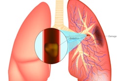
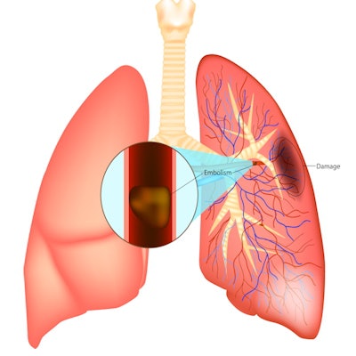
Acute PE is a potentially lethal condition, and timely diagnosis and treatment is key to optimizing outcomes, Pannenbecker said. CTPA is the reference standard for diagnostic workup of suspected PE; dual-energy CT offers even more benefit because it produces spectral information, she noted. PCCT is a multienergy technique that offers this spectral information but at a lower radiation dose, according to Pannenbecker.
The team sought to evaluate objective and subjective image quality of an ultra-low contrast medium and radiation dose CT pulmonary angiography (CTPA) protocol for diagnosis of acute PE. They used a photon-counting detector CT scanner and compared the results to a conventional energy-integrating detector (EID) based dual-energy CTPA protocol.
The study included 32 photon-counting detector CTPA exams using 25 ml of contrast and 2.5 mGy*cm of CT dose index volume (CTDIvol) and 32 conventional CT exams using 50 ml of contrast and 5.1 mGy*cm of CTDIvol. Four readers evaluated the images; the team compared pulmonary artery CT attenuation, signal-to-noise ratio, contrast-to-noise ratio, and subjective image quality at 60 keV using virtual monoenergetic imaging (VMI) and polychromatic standard reconstructions, Pannenbecker said.
The researchers found that CT attenuation, signal-to-noise ratio, and contrast-to-noise ratio were significantly higher in the conventional CT exam group (p < 0.001), and that the four readers rated subjective image quality of 60 keV photon-counting detector scans as excellent or good in 94.4 % of the photon-counting detector CT images compared to 84.4 % of conventional scans.
| Performance comparison for identifying pulmonary embolism, conventional CT, and PCCT | |||
| Measure | Conventional CT | PCCT | p-value |
| Dose length product (mGy*cm) | 181.7 | 80 | < 0.001 |
| Effective dose (mSv) | 3.3 | 1.4 | < 0.001 |
| Size specific dose estimate (mGy) | 5.7 | 3.1 | < 0.001 |
"Even though we halved the contrast medium dose and radiation dose compared to standard dual-energy [conventional CT] protocol, [we maintained] good to excellent diagnostic imaging quality in nearly 100% of cases," she concluded.