Sunday, November 27 | 9:00 a.m.-10:00 a.m. | S1-SSCH01-5 | Room E351
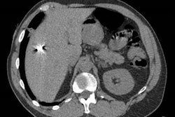
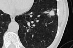
The effects of COVID-19 on patients' lungs are evident on CT, even a year after initial infection, according to this Sunday morning presentation.Presenter Dr. Martine Remy-Jardin, PhD, of the University Centre of Lille in France will share results from a study that included 79 patients who were hospitalized for COVID-19 pneumonia between March 2020 and April 2021. Patients were followed-up between six and 12 months with a dual-energy CT angiography exam and review of CT lung perfusion images.
COVID-19 patients continued to show abnormalities in their lungs in the year following hospitalization, the team found. CT showed acute pulmonary embolism features in two patients (2.5%) and focal chronic thromboembolic disease in three (3.8%), it also showed residual post-COVID-19 lung infiltration involving 4.7% of lung volume in 85% of patients. Radiologist readers rated lung perfusion as abnormal in 87.4% of study participants: These abnormal findings included patchy defects, areas of nonsystematized hypoperfusion, and pulmonary embolism-like defects.
The findings could give clinicians a better understanding about how COVID-19 affects people long-term.
"[Later] follow-up showed CT features of acute and chronic pulmonary embolism but also two types of perfusion abnormalities suggestive of persistent hypercoagulability as well as unresolved sequelae of the widespread [cerebral small vessel disease] described in the acute phase of the disease," the group concluded.


![Images show the pectoralis muscles of a healthy male individual who never smoked (age, 66 years; height, 178 cm; body mass index [BMI, calculated as weight in kilograms divided by height in meters squared], 28.4; number of cigarette pack-years, 0; forced expiratory volume in 1 second [FEV1], 97.6% predicted; FEV1: forced vital capacity [FVC] ratio, 0.71; pectoralis muscle area [PMA], 59.4 cm2; pectoralis muscle volume [PMV], 764 cm3) and a male individual with a smoking history and chronic obstructive pulmonary disorder (COPD) (age, 66 years; height, 178 cm; BMI, 27.5; number of cigarette pack-years, 43.2, FEV1, 48% predicted; FEV1:FVC, 0.56; PMA, 35 cm2; PMV, 480.8 cm3) from the Canadian Cohort Obstructive Lung Disease (i.e., CanCOLD) study. The CT image is shown in the axial plane. The PMV is automatically extracted using the developed deep learning model and overlayed onto the lungs for visual clarity.](https://img.auntminnie.com/mindful/smg/workspaces/default/uploads/2026/03/genkin.25LqljVF0y.jpg?auto=format%2Ccompress&crop=focalpoint&fit=crop&h=100&q=70&w=100)


![Images show the pectoralis muscles of a healthy male individual who never smoked (age, 66 years; height, 178 cm; body mass index [BMI, calculated as weight in kilograms divided by height in meters squared], 28.4; number of cigarette pack-years, 0; forced expiratory volume in 1 second [FEV1], 97.6% predicted; FEV1: forced vital capacity [FVC] ratio, 0.71; pectoralis muscle area [PMA], 59.4 cm2; pectoralis muscle volume [PMV], 764 cm3) and a male individual with a smoking history and chronic obstructive pulmonary disorder (COPD) (age, 66 years; height, 178 cm; BMI, 27.5; number of cigarette pack-years, 43.2, FEV1, 48% predicted; FEV1:FVC, 0.56; PMA, 35 cm2; PMV, 480.8 cm3) from the Canadian Cohort Obstructive Lung Disease (i.e., CanCOLD) study. The CT image is shown in the axial plane. The PMV is automatically extracted using the developed deep learning model and overlayed onto the lungs for visual clarity.](https://img.auntminnie.com/mindful/smg/workspaces/default/uploads/2026/03/genkin.25LqljVF0y.jpg?auto=format%2Ccompress&crop=focalpoint&fit=crop&h=112&q=70&w=112)


