Neuroscientists used electroencephalography (EEG) readings on an 87-year-old epileptic man who was dying from a heart attack to discover brain activity similar to memory "flashbacks," according to a report published February 23 in the Daily Mail.
"This supports a theory known as 'life recall' -- that we relive our entire life in the space of seconds like a flash of lightning just prior to death," the report said. "In fact, the brain may remain active and coordinated during and after the transition to death and may even be programmed to 'orchestrate the whole ordeal.' "
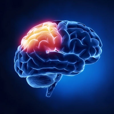
The patient was admitted to the Vancouver General Hospital in British Columbia and was under the care of Dr. Ajmal Zemmar, who acquired CT images and EEG readings. The EEG readings measured 900 seconds of brain activity before and after the man died, according to the Daily Mail.
"Just before and after the heart stopped working, we saw changes in a specific band of neural oscillations, so-called gamma oscillations, but also in others such as delta, theta, alpha, and beta oscillations," Zemmar said in the report.
Brain waves of this sort make up cognitive functions such as "concentrating, dreaming, meditation, memory retrieval, information processing, and conscious perception, just like those associated with memory flashbacks," the Daily Mail said.