
Tuesday, November 30 | 9:30 a.m.-10:30 a.m. | SSPH09-1 | Room TBA
Radiation dose for a novel spiral breast CT scanner can be determined by measuring breast volume, according to this presentation.And the radiation dose may be lower than the dose limit for screening mammography, presenter Sojin Shim of the University Hospital in Zurich, Switzerland, and colleagues noted. Shim is a doctoral candidate in diagnostic and interventional radiology.
"[We] discovered that, with the current exposure setup for a medium-large breast, the opportunistic and diagnostic [spiral] breast CT scan results in mean glandular dose values lower than the dose limit for a screening mammography on a medium-large breast mandated by the European commission," the researchers wrote in the study abstract.
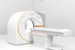
Spiral breast CT is a relatively new technique for breast imaging. Shim's team sought to estimate the level of radiation dose women receive during this type of exam and included 1,037 patients in their research.
Each of the women underwent spiral breast CT; the images were segmented using image processing algorithms developed by the group into one of four categories: adipose tissue, glandular tissue, skin, and pectoralis major muscle. The group measured breast and skin volume and breast density on the segmented images, and calculated the following five dose values:
- Mean absorbed dose in the breast
- Mean glandular dose
- Mean skin dose
- Max 5% glandular dose
- Max 5% skin dose
These dose parameters were then matched to breast features.
Breast volume had the best correlation with dose parameters, Shim's team found. Seventy percent of breasts had a volume of more than 390 cm3, and for these, the average dose values for the five categories were 6.56 mGy, 6.25 mGy, 10.69 mGy, 10.29 mGy, and 19.27 mGy, respectively.
"We established for the first time a radiation dose estimation model for spiral breast CT scans that requires only a simple breast volume measurement," Shim and colleagues wrote.


![Axial images from unenhanced calcium score cardiac CT (left) and curved planar reformation images from CT angiography (right) show that higher long-term exposure to air pollution is associated with greater coronary artery calcium and more obstructive coronary artery disease (CAD). Top row: Images in a 68-year-old male patient with higher 10-year mean ambient air pollution exposure (7.9 μg/m3 for particulate matter measuring ≤2.5 μm in diameter [PM2.5] and 17.4 parts per billion [ppb] for NO2) with extensive CAD (coronary artery calcium score [CACS] >1,000 and obstructive CAD [≥70% diameter stenosis]). Bottom row: Images in a 57-year-old female patient with lower 10-year mean ambient air pollution exposure (6.3 μg/m3 for PM2.5 and 4.6 ppb for NO2) with no CAD (CACS = 0 and no obstructive stenosis).](https://img.auntminnie.com/mindful/smg/workspaces/default/uploads/2026/06/hanneman.r6SMLzkezo.png?auto=format%2Ccompress&dpr=2&fit=crop&h=167&q=70&w=250)


