
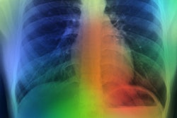
Thoracic CT in patients who are slated to undergo abdominal or pelvic surgery can help clinicians better predict which may require mechanical ventilation after the procedure, according to a study published September 2 in the American Journal of Roentgenology.
The study findings could help surgeons better plan for potential postsurgery risks, wrote a team led by Dr. Arzu Canan of the University of Texas Southwestern Medical Center in Dallas.
"Many patients undergo thoracic CT before abdominal or pelvic surgery; the CT findings may complement preoperative clinical risk factors," Canan and colleagues noted.
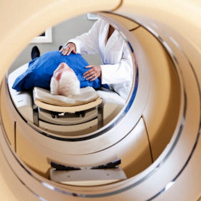
The study included 165 patients who underwent thoracic CT before abdominal or pelvic surgery that required general endotracheal tube anesthesia. Of these, 42 required postoperative mechanical ventilation and 123 did not.
Canan and colleagues found that on preoperative CT, predictors of postoperative mechanical ventilation included the following:
- Bronchial wall thickening (odds ratio, 4.8)
- Pericardial effusion (odds ratio, 5.3)
- Shorter lung height (odds ratio, 0.8 per cm increase)
- Greater anteroposterior chest diameter (odds ratio, 1.2 per cm increase)
"Bronchial wall thickening was the only qualitative parameter involving the lung parenchyma that differed between the case and control groups ... with significantly increased need for postoperative mechanical ventilation in patients with [it]," Canan and colleagues concluded.


![Images show the pectoralis muscles of a healthy male individual who never smoked (age, 66 years; height, 178 cm; body mass index [BMI, calculated as weight in kilograms divided by height in meters squared], 28.4; number of cigarette pack-years, 0; forced expiratory volume in 1 second [FEV1], 97.6% predicted; FEV1: forced vital capacity [FVC] ratio, 0.71; pectoralis muscle area [PMA], 59.4 cm2; pectoralis muscle volume [PMV], 764 cm3) and a male individual with a smoking history and chronic obstructive pulmonary disorder (COPD) (age, 66 years; height, 178 cm; BMI, 27.5; number of cigarette pack-years, 43.2, FEV1, 48% predicted; FEV1:FVC, 0.56; PMA, 35 cm2; PMV, 480.8 cm3) from the Canadian Cohort Obstructive Lung Disease (i.e., CanCOLD) study. The CT image is shown in the axial plane. The PMV is automatically extracted using the developed deep learning model and overlayed onto the lungs for visual clarity.](https://img.auntminnie.com/mindful/smg/workspaces/default/uploads/2026/03/genkin.25LqljVF0y.jpg?auto=format%2Ccompress&crop=focalpoint&fit=crop&h=100&q=70&w=100)


![Images show the pectoralis muscles of a healthy male individual who never smoked (age, 66 years; height, 178 cm; body mass index [BMI, calculated as weight in kilograms divided by height in meters squared], 28.4; number of cigarette pack-years, 0; forced expiratory volume in 1 second [FEV1], 97.6% predicted; FEV1: forced vital capacity [FVC] ratio, 0.71; pectoralis muscle area [PMA], 59.4 cm2; pectoralis muscle volume [PMV], 764 cm3) and a male individual with a smoking history and chronic obstructive pulmonary disorder (COPD) (age, 66 years; height, 178 cm; BMI, 27.5; number of cigarette pack-years, 43.2, FEV1, 48% predicted; FEV1:FVC, 0.56; PMA, 35 cm2; PMV, 480.8 cm3) from the Canadian Cohort Obstructive Lung Disease (i.e., CanCOLD) study. The CT image is shown in the axial plane. The PMV is automatically extracted using the developed deep learning model and overlayed onto the lungs for visual clarity.](https://img.auntminnie.com/mindful/smg/workspaces/default/uploads/2026/03/genkin.25LqljVF0y.jpg?auto=format%2Ccompress&crop=focalpoint&fit=crop&h=112&q=70&w=112)


