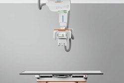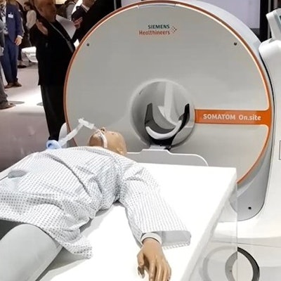
This year's winner of the Minnies award for Best New Radiology Device is the Somatom On.site portable CT scanner from Siemens Healthineers. On.site represents a departure from traditional CT instrumentation -- it mounts a dedicated head CT scanner on a mobile gantry that can be wheeled to the patient's bedside.
Siemens developed the system to enable clinicians to make CT technology more available in areas like the intensive care unit, where patients might need closer monitoring. Siemens executives Doug Ryan, vice president of the CT business line for Siemens Healthineers North America, and Philipp Fischer, head of CT at Siemens Healthineers, spoke with AuntMinnie.com Editor-in-Chief Brian Casey about the product.
Now in their 21st year, the Minnies awards are AuntMinnie.com's annual event recognizing excellence in radiology, with over 200 candidates competing in 15 categories, ranging from Most Influential Radiology Researcher to Radiology Image of the Year.
Minnies candidates are nominated by AuntMinnie.com members, with winners selected by an expert panel of radiology luminaries in two rounds of voting. Winners are recognized each year at the annual RSNA meeting. A full list of winners in the 2020 edition of the Minnies is available on AuntMinnie.com.