
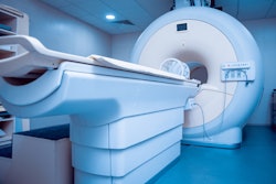
In an October 5 press conference, President Donald Trump's physician declined to discuss the findings of medical imaging exams the president received as part of his care for COVID-19 at Walter Reed National Military Medical Center in Bethesda, MD.
During the presser, Dr. Sean Conley said the president's healthcare team has performed "routine standard imaging" but that he was not at liberty to discuss the findings. When pressed further, Conley remained tight-lipped about what types of imaging the president has undergone and any potential findings, citing HIPAA restrictions.
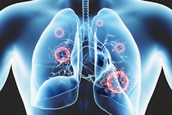
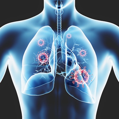
"So you're actively not telling us what those lung scans show, just to be clear?" a reporter asked.
To which Conley responded, "There are HIPAA rules and regulations that restrict me in sharing certain things for his safety and his own health -- and reasons."
Conley also invoked HIPAA when asked about any abnormal findings on blood work and whether the president was taking a blood thinner. However, the care team readily shared other health information, including the president's temperature, blood pressure, respiratory rate, blood oxygen saturation, and use of both Remdesivir and dexamethasone.
The press conference occurred shortly after the president announced on Twitter that he would be discharged from Walter Reed later today. Conley said President Trump met their discharge criteria to return to the White House but isn't yet in the clear.
"Though he may not entirely be out of the woods yet, the team and I agree that all our evaluations -- and most importantly his clinical status -- support the president's safe return home where he'll be surrounded by world-class medical care 24/7," Conley said.


![Images show the pectoralis muscles of a healthy male individual who never smoked (age, 66 years; height, 178 cm; body mass index [BMI, calculated as weight in kilograms divided by height in meters squared], 28.4; number of cigarette pack-years, 0; forced expiratory volume in 1 second [FEV1], 97.6% predicted; FEV1: forced vital capacity [FVC] ratio, 0.71; pectoralis muscle area [PMA], 59.4 cm2; pectoralis muscle volume [PMV], 764 cm3) and a male individual with a smoking history and chronic obstructive pulmonary disorder (COPD) (age, 66 years; height, 178 cm; BMI, 27.5; number of cigarette pack-years, 43.2, FEV1, 48% predicted; FEV1:FVC, 0.56; PMA, 35 cm2; PMV, 480.8 cm3) from the Canadian Cohort Obstructive Lung Disease (i.e., CanCOLD) study. The CT image is shown in the axial plane. The PMV is automatically extracted using the developed deep learning model and overlayed onto the lungs for visual clarity.](https://img.auntminnie.com/mindful/smg/workspaces/default/uploads/2026/03/genkin.25LqljVF0y.jpg?auto=format%2Ccompress&crop=focalpoint&fit=crop&h=100&q=70&w=100)


![Images show the pectoralis muscles of a healthy male individual who never smoked (age, 66 years; height, 178 cm; body mass index [BMI, calculated as weight in kilograms divided by height in meters squared], 28.4; number of cigarette pack-years, 0; forced expiratory volume in 1 second [FEV1], 97.6% predicted; FEV1: forced vital capacity [FVC] ratio, 0.71; pectoralis muscle area [PMA], 59.4 cm2; pectoralis muscle volume [PMV], 764 cm3) and a male individual with a smoking history and chronic obstructive pulmonary disorder (COPD) (age, 66 years; height, 178 cm; BMI, 27.5; number of cigarette pack-years, 43.2, FEV1, 48% predicted; FEV1:FVC, 0.56; PMA, 35 cm2; PMV, 480.8 cm3) from the Canadian Cohort Obstructive Lung Disease (i.e., CanCOLD) study. The CT image is shown in the axial plane. The PMV is automatically extracted using the developed deep learning model and overlayed onto the lungs for visual clarity.](https://img.auntminnie.com/mindful/smg/workspaces/default/uploads/2026/03/genkin.25LqljVF0y.jpg?auto=format%2Ccompress&crop=focalpoint&fit=crop&h=112&q=70&w=112)


