Dutch researchers have released images acquired using micro-CT that depict a 6-week-old ectopic pregnancy. The case study was published August 11 in Radiology.
"Micro-CT provides an unprecedented look into early human development," wrote Dr. Yousif Dawood and Dr. Bernadette de Bakker of the University of Amsterdam.
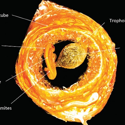
 An intact ectopic pregnancy in the fallopian tube was imaged by contrast-enhanced micro-CT at 3-µm isotropic resolution, revealing a 6-week-gestation human embryo 3 mm in length. The embryo and its yolk sac are completely surrounded by trophoblast (developing placenta) and the fallopian tube. The embryo is shown from the dorsal side, with the developing neural tube facing the viewer. Image courtesy of the RSNA.
A woman who presented with abdominal pain and a positive pregnancy test underwent transvaginal ultrasound, which showed an ectopic pregnancy in the left fallopian tube. The tube was removed via laparoscopy and immersed in an iodine and potassium iodide solution for two days before being scanned with micro-CT at 3-µm resolution; total scanning time was 45 minutes.
 The embryo is shown from the ventral side, with the developing heart tube facing the viewer. Various organs are denoted. Image courtesy of the RSNA.
"Comparison to images in the three-dimensional atlas of human embryology showed full resemblance with a stage 12 embryo, corresponding with 28 developmental days (6 pregnancy weeks)," the team wrote.