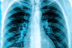
The World Health Organization (WHO) published rapid guidance on July 30 in Radiology regarding the use of chest imaging for the diagnosis and management of patients with COVID-19.
An international team of authors developed the guide over two months using online meetings and a process of rapid systematic reviews to assess the acceptability, feasibility, and impact of chest radiography, chest CT, and lung ultrasound for COVID-19 imaging.
The guide includes three recommendations for COVID-19 diagnosis:
- Chest imaging is not suggested to diagnose COVID-19 in asymptomatic patients.
- Chest imaging is not suggested to diagnose COVID-19 in symptomatic patients when reverse transcription polymerase chain reaction (RT-PCR) testing is available and timely.
- Chest imaging is suggested to diagnose COVID-19 in symptomatic patients when RT-PCR testing is not available or has delayed results, or when an initial RT-PCR test is negative but there is still high clinical suspicion of COVID-19.
The guidance also contains four recommendations for using imaging for patient management:
- Chest imaging is suggested to help clinicians decide whether to admit or discharge patients with mild COVID-19 symptoms.
- Chest imaging is suggested to help clinicians decide whether to admit patients with moderate-to-severe COVID-19 symptoms to a regular ward or to an intensive care unit.
- Chest imaging is suggested to help inform the therapeutic management of hospitalized patients with moderate-to-severe COVID-19 symptoms.
- Chest imaging is not suggested to help inform discharge decisions for hospitalized patients whose symptoms have resolved.
The authors cautioned the recommendations are conditional and based on expert opinions and low or very low certainty evidence from 28 studies. The recommendations are also for chest imaging in general -- not for specific modalities.
"While there is accumulating evidence about typical findings with each imaging modality, evidence about comparative diagnostic and prognostic value of the different modalities is still lacking," the authors wrote.