
A Philips Healthcare team has converted shipping containers into offsite COVID-19 radiology suites in the Philippines.
The repurposed containers, known as imaging cabins, contain either CT or x-ray systems, according to the vendor. Once created, the cabins can be transported and placed wherever they are needed, such as within a hospital, on hospital grounds, or within a community, Philips said.
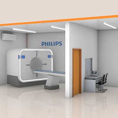
An illustration of a CT cabin. Image courtesy of Royal Philips.Radiology departments can use the cabins to perform diagnostic imaging procedures with minimal or no patient contact, according to Philips. The cabins also have a lead shield and ultraviolet lamps designed to help prevent stray radiation and assist with sterilization.
The suites are also equipped with a computer for quick image analysis and can be linked to the hospital IT network for remote reading as well, according to the firm.


![Images show the pectoralis muscles of a healthy male individual who never smoked (age, 66 years; height, 178 cm; body mass index [BMI, calculated as weight in kilograms divided by height in meters squared], 28.4; number of cigarette pack-years, 0; forced expiratory volume in 1 second [FEV1], 97.6% predicted; FEV1: forced vital capacity [FVC] ratio, 0.71; pectoralis muscle area [PMA], 59.4 cm2; pectoralis muscle volume [PMV], 764 cm3) and a male individual with a smoking history and chronic obstructive pulmonary disorder (COPD) (age, 66 years; height, 178 cm; BMI, 27.5; number of cigarette pack-years, 43.2, FEV1, 48% predicted; FEV1:FVC, 0.56; PMA, 35 cm2; PMV, 480.8 cm3) from the Canadian Cohort Obstructive Lung Disease (i.e., CanCOLD) study. The CT image is shown in the axial plane. The PMV is automatically extracted using the developed deep learning model and overlayed onto the lungs for visual clarity.](https://img.auntminnie.com/mindful/smg/workspaces/default/uploads/2026/03/genkin.25LqljVF0y.jpg?auto=format%2Ccompress&crop=focalpoint&fit=crop&h=112&q=70&w=112)


