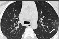
Although the clinical presentation of various lung diseases in children can show significant overlap, there are emerging characteristic imaging findings -- such as the halo sign for COVID-19 on chest CT -- that can help radiologists make more accurate diagnoses, according to a study published April 29 in the American Journal of Roentgenology.
Clinical symptoms of pediatric lung disorders like severe acute respiratory syndrome (SARS), swine-origin influenza A (H1N1), Middle East respiratory syndrome (MERS), electronic-cigarette (e-cigarette) or vaping-associated lung injury (EVALI), and the novel coronavirus disease (COVID-19) may be nonspecific, but distinguishing imaging findings are becoming more clear, wrote a team led by Dr. Alexandra Foust of Boston Children's Hospital.
"Although there are some overlapping imaging features of these disorders, careful evaluation of the distribution, lung zone preference, and symmetry of the abnormalities with an eye for a few unique differentiating imaging features, such as the halo sign seen in COVID-19 and subpleural sparing and the atoll sign seen in EVALI, can allow the radiologist to offer a narrower differential diagnosis in pediatric patients," Foust and colleagues wrote.
For example, in 20 chest CTs of pediatric patients with COVID-19, the group found that abnormalities included subpleural lesions (100%), unilateral (30%) or bilateral (50%) pulmonary lesions, ground-glass opacities (60%), and the halo sign (50%), according to the researchers.
These distinguishing imaging characteristics can help clinicians better diagnose pediatric patients, the group concluded.
"It is essential for radiologists to have a clear understanding of the characteristic imaging appearances of these lung disorders in pediatric patients to ensure optimal patient care," it wrote.