Dear AuntMinnie Member,
In what's become an annual tradition, the Medicare Payment Advisory Commission (MedPAC) has included a warning about the growth of imaging utilization in its annual report released today to the U.S. Congress.
The problem is that imaging utilization has actually declined 7% since 2009 -- a fact that MedPAC acknowledges in the new report. But instead of highlighting what should be good news (at least to public policymakers), the commission chose instead to warn that imaging utilization is now higher than it was in 2000 -- nearly two decades ago.
MedPAC's motivation for continuing to warn about a problem that doesn't seem to exist anymore is unclear; perhaps commission members have a nostalgic urge to relive the good old days. The danger to radiology is that the commission's quixotic warnings could be taken seriously by policymakers who don't know better and continue to look for cuts to radiology reimbursement.
Get the rest of the story by clicking here, or visit our Imaging Leaders Community at leaders.auntminnie.com.
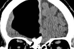
CT scan reveals air pocket in head
Meanwhile, we're highlighting one of the more unusual cases we've run across recently in our CT Community. A man in Northern Ireland presented to a local hospital complaining of unsteadiness and recurrent falls. His healthcare providers ordered a brain CT scan to help figure out what was wrong.
The images revealed a large pocket of air in the man's right frontal lobe, as well as an osteoma nearby. The osteoma had apparently eroded the bone plate near the nasal cavity, allowing air to enter the right frontal lobe and create the air pocket. Learn more about this fascinating case by clicking here, or visit our CT Community at ct.auntminnie.com.
Does DBT change 2D interpretation?
Finally, visit our Women's Imaging Community for a new article that examines whether experience with digital breast tomosynthesis (DBT) changes the way breast imagers read 2D mammograms. Researchers found that after breast specialists read DBT images, they had higher recall rates when interpreting 2D images -- but also higher cancer detection rates. Learn what might be going on by clicking here, or visit our Women's Imaging Community at women.auntminnie.com.