Collecting bone mineral density measurements from routine CT scans of elderly individuals could help predict their risk of experiencing an osteoporotic fracture, according to a study published online February 5 in the Journal of Bone and Mineral Research.
Researchers from the University of Wisconsin-Madison collected bone density values from the thoracoabdominal CT scans of patients who were at least 65 years old and who underwent CT exams in 2003 for clinical indications other than osteoporosis. Out of 507 patients, 114 turned out to have a fracture within 10 years of the initial CT exam.
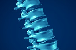
After analyzing the data, the group found that trabecular attenuation in the first lumbar vertebra of less than 90 Hounsfield units (HU) was associated with an increased risk of fracture within six years (p < 0.001). What's more, an increase of 10 units in trabecular attenuation in the same region was associated with a notable decrease in fracture risk after a CT exam.
"CT scans are commonly performed in older adults for a wide variety of reasons," senior author Dr. Perry Pickhardt said in a statement. "The rich bone data embedded in these scans is often ignored, but can and should be harnessed for opportunistic screening for fracture risk."