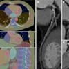
Medical artificial intelligence (AI) firm DeepRadiology has introduced its CT head AI software and is exhibiting at RSNA 2017 in Chicago.
The software detects significant pathologies in head CT scans with error rates better than published rates for radiologists, according to the company.
DeepRadiology created the software using artificial intelligence (AI) techniques, the knowledge contained in all major radiology textbooks on the subject, and the experience of reviewing more than 9 million CT scan images.
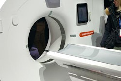
The firm has developed similar systems for other CT scan types and images from plain radiographs, MRI, ultrasound, mammography, and nuclear medicine.


![Images show the pectoralis muscles of a healthy male individual who never smoked (age, 66 years; height, 178 cm; body mass index [BMI, calculated as weight in kilograms divided by height in meters squared], 28.4; number of cigarette pack-years, 0; forced expiratory volume in 1 second [FEV1], 97.6% predicted; FEV1: forced vital capacity [FVC] ratio, 0.71; pectoralis muscle area [PMA], 59.4 cm2; pectoralis muscle volume [PMV], 764 cm3) and a male individual with a smoking history and chronic obstructive pulmonary disorder (COPD) (age, 66 years; height, 178 cm; BMI, 27.5; number of cigarette pack-years, 43.2, FEV1, 48% predicted; FEV1:FVC, 0.56; PMA, 35 cm2; PMV, 480.8 cm3) from the Canadian Cohort Obstructive Lung Disease (i.e., CanCOLD) study. The CT image is shown in the axial plane. The PMV is automatically extracted using the developed deep learning model and overlayed onto the lungs for visual clarity.](https://img.auntminnie.com/mindful/smg/workspaces/default/uploads/2026/03/genkin.25LqljVF0y.jpg?auto=format%2Ccompress&crop=focalpoint&fit=crop&h=112&q=70&w=112)

