Dear CT Insider,
Dose alerts are designed to warn CT scanner operators when a protocol might exceed predetermined radiation dose limits. Annoying at times, they are nevertheless required equipment on today's scanners, thanks to a U.S. government initiative that will soon levy reimbursement penalties on scans completed without them.
But how good are the alerts in practice? Do they really prevent radiation damage to the skin, which is the reason they were developed? Dr. Aaron Sodickson, PhD, from Brigham and Women's Hospital in Boston wondered how much help the alerts provided in the clinical setting, and who might still be at risk when the warnings fail -- which is often. The results, provided in our Insider Exclusive, might surprise you.
Of course, by the time a scan gets to the dose alert stage, something important might have been missed along the way. Radiation dose management on the enterprise level is actually fairly straightforward, but it comes with a lot of moving parts and requires constant vigilance. Learn how one medical physicist approaches this daunting task by clicking here.
A key target of dose optimization efforts over the years has been the head, which is good news considering that a new study shows an avalanche of head CT exams being performed in children as a result of sports-related injuries. How many? Find out here. On the other hand, a study from New York found that common CT exams are being ordered appropriately, potentially tamping down demands for further imaging cuts.
Another new study reveals disturbing information about coronary CT angiography (CCTA) and radiation risk: Researchers have found measurable DNA damage following some CCTA scans. Fortunately, however, the damage doesn't apply to most scans performed today.
Researchers at five University of California schools took a broad approach to evaluating radiation doses in practice, measuring dose in tens of thousands of CT exams given to adults and children to see how well they were doing in terms of optimizing dose. See what they found here.
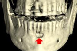
Armed with new analytical tools, CT providers are revealing new information about imaging findings that allow some conditions to be treated noninvasively. Such is the case with parathyroid lesions, where adjusting the protocols just a bit can reveal three distinct lesion types requiring different treatment approaches. Get the rest of the story here.
Finally, a group at Massachusetts General Hospital in Boston is saving money by printing 3D hearts for surgical planning and patient education.
Get all the other stories in this issue by scrolling through the links below. And for all the latest news in CT imaging, you'll want to stick by your CT Community.