Two new studies presented this week at the Society of NeuroInterventional Surgery (SNIS) meeting in San Francisco concluded that mobile stroke treatment units -- ambulances equipped with CT scanners -- can significantly reduce the time needed to diagnose and treat stroke patients, likely improving survival and boosting chances of recovery.
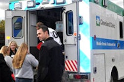
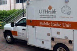
Four mobile stroke units are currently in use: two in Germany and two in the U.S., in Cleveland and Houston. Each unit carries a conebeam CT scanner and other diagnostic tools, along with telemedicine capabilities and intravenous tissue plasminogen activator (tPA) for onboard treatment of ischemic stroke once confirmed at CT.
Because even a few minutes' delay can result in large-scale brain loss, the principal goal of the stroke units is to diagnose patients and determine the course of treatment as quickly as possible, said Dr. Shazam Hussain, head of the Cleveland Clinic Stroke Program, in a statement accompanying the two SNIS presentations.
In the first SNIS study, Cleveland Clinic researchers discussed how patients who received IV tPA from mobile stroke units showed improved blood flow restoration rates for emergent large-vessel occlusion (clots) strokes that were treated in the hour following symptom onset.
In another Cleveland Clinic study, patients who received intra-arterial therapy in mobile stroke units for large-vessel occlusion strokes saw decreased time spent at many points in the diagnostic/treatment pathway, including ambulance dispatch to door (19 minutes with a mobile unit versus 31 minutes); door to initial CT scan (12 minutes versus 32 minutes); and CT to intra-arterial therapy (82 minutes versus 165 minutes). All differences were statistically significant.