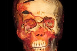
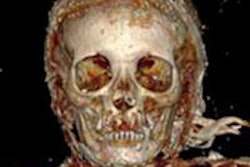
Researchers from the Buffalo Museum of Science in Buffalo, NY, have conducted a virtual autopsy on a South American mummy using 3D CT, the museum said.
As part of the museum's "Mummies of the World" exhibition, Dr. Peter Loud of Roswell Park Cancer Institute and Heather Gill-Frerking, PhD, of American Exhibitions imaged the mummy and discovered she was a 2-year-old girl. The scan also found no evidence of disease or trauma to the skeleton.
The mummy has been part of the Buffalo Museum of Science's collection for more than 100 years, according to the museum.


![Images show the pectoralis muscles of a healthy male individual who never smoked (age, 66 years; height, 178 cm; body mass index [BMI, calculated as weight in kilograms divided by height in meters squared], 28.4; number of cigarette pack-years, 0; forced expiratory volume in 1 second [FEV1], 97.6% predicted; FEV1: forced vital capacity [FVC] ratio, 0.71; pectoralis muscle area [PMA], 59.4 cm2; pectoralis muscle volume [PMV], 764 cm3) and a male individual with a smoking history and chronic obstructive pulmonary disorder (COPD) (age, 66 years; height, 178 cm; BMI, 27.5; number of cigarette pack-years, 43.2, FEV1, 48% predicted; FEV1:FVC, 0.56; PMA, 35 cm2; PMV, 480.8 cm3) from the Canadian Cohort Obstructive Lung Disease (i.e., CanCOLD) study. The CT image is shown in the axial plane. The PMV is automatically extracted using the developed deep learning model and overlayed onto the lungs for visual clarity.](https://img.auntminnie.com/mindful/smg/workspaces/default/uploads/2026/03/genkin.25LqljVF0y.jpg?auto=format%2Ccompress&crop=focalpoint&fit=crop&h=112&q=70&w=112)


