SAN FRANCISCO - Dual-energy virtual colonoscopy may be able to replace several other exams in patients with colorectal cancer -- all while streamlining staging, surgical planning, and the search for metastases. Preliminary research results, presented last week at the International Society for Computed Tomography (ISCT) annual meeting, suggest high accuracy and some unique capabilities for the technique.
Dual-energy CT (DECT) images, acquired on both single- and dual-source scanners, have already earned high marks in several imaging applications, including suspected pulmonary embolism and enhancing renal masses.
Although development of the technique in virtual colonoscopy (also known as CT colonography or CTC) is still in the early stages, it is primed for greater popularity due to its ability to predict tumor histology based on contrast uptake, according to Dr. Anno Graser from the University of Munich.
"We provide the true one-stop examination," Graser said. With DECT, "two images, a high-kV and a low-kV image, can be combined into a weighted-average image that is very similar to a 120-kVp image. We can subtract out the iodine and generate an iodine-only image, we can generate virtual unenhanced images, and we can superimpose them onto each other."
When to use it? Everybody who is diagnosed with colorectal cancer needs staging CT and also colonoscopy, Graser said. But dual-energy CTC has the potential to provide all the necessary information in a single scan, including:
- Local tumor staging
- Nodal metastases staging
- Search for synchronous polyps and cancers
- Assessment of organ metastases
"If you combine CT colonography with dual-energy techniques, you can get information about lesion histology, and this might have the potential to differentiate adenomas from hyperplastic polyps," Graser said.
The technique can be used to determine the contrast media uptake of the polyp, as well as the baseline density of the lesion, directly measuring enhancement in HU and measuring the iodine in the lesion in grams per mL.
So-called DECTC does come with one limitation regular CTC practitioners won't appreciate. "You cannot use this to subtract tagged fluid from the colon, because if you subtract the iodine you're going to be left with water, and water is just as bad as iodine-tagged fluid in the protocol," Graser said. Also, a posterior/anterior topogram is needed to ensure that the patient is centered in the scan field due to the smaller size of one of the detectors in the dual-source CT scanner.
Images are acquired prone and supine as in a normal CTC exam on a dual-source scanner (Somatom Flash, Siemens Healthcare), following cathartic bowel cleansing with 2 L of polyethylene glycol, automated insufflation of CO2 (ProtoCO2l, Bracco Diagnostics), and injection of 1.3 mL per kg of body weight of contrast media (Iomeron 400, Bracco) for the supine scan.
The unenhanced prone acquisition is acquired using a section width of 128 x 0.6 mm, 120 kVp, and 30 mAs. The dual-energy acquisition is a standard abdominal protocol: 100/140 kV with a tin filter, 300 mAs, 32 x 0.6 collimation, and 0.6 pitch, Graser said, reconstructed in 5-mm increments and viewed on a PACS workstation using weighted-average images.
 |
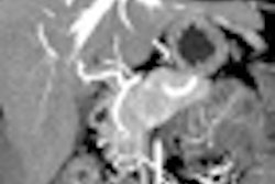
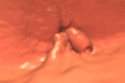
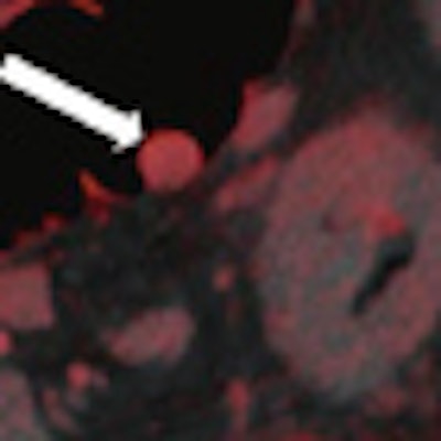
| Images are of a 65-year-old male patient who underwent a single acquisition at dual-source CTC. Above, a stenosing carcinoma was seen in the proximal sigmoid colon in 3D endoluminal views (left, middle) and 2D view (right). Below, an 18-mm colorectal adenoma was also found in the proximal sigmoid colon, and also appears to be enhancing. Bottom image shows an enhancing liver metastasis in the same patient. Virtual unenhanced (grayscale) images are created by electronically subtracting the contrast media. All images courtesy of Dr. Anno Graser. |
 |
 |
The technique is ideal for examining the entire colon in patients with a stenosing colorectal cancer lesion, providing additional insight into the region proximal to the stenosing cancer as well, he said.
The Munich group has evaluated the first results of an ongoing trial including 25 patients (mean age, 63 ± 8 years) who underwent a full cathartic bowel prep prior to DSCTC on the day of surgery.
"Normally for colon cancer resection they wouldn't perform a full bowel prep anymore, but [surgeons] still like it if it's there," Graser said. The results were correlated to intraoperative findings, colonoscopy, and histopathology of the lesions.
Preliminary results included the following:
The primary tumor stage was accurate in 24 of 25 patients (96%).
Synchronous cancers were found in two patients (8%).
Synchronous adenomas were identified in nine of 25 patients (36%). Five patients had one adenoma, two patients had two adenomas, and one patient had three adenomas.
The size of adenomas ranged from 6 to 27 mm.
The mean enhancement of adenomas was 55 ± 27 HU.
Hyperplastic lesions were indentified in four patients.
The mean enhancement of hyperplastic lesions was 13 ± 10 HU.
"Dual-energy CTC is a unique method to characterize colonic lesions based on their enhancement" in colorectal cancer patients, Graser said. "High-resolution imaging of the CO2-distended large bowel" offers "true one-stop imaging of the large bowel, which is why the surgeons really came to love this."
The method provides "local tumor staging, lymph node staging, distant metastases staging, and the localization of synchronous polyps and cancers," he said.