In a new study that tested phantoms as well as patients, a commercially available bismuth shield for the thyroid gland substantially reduced CT radiation dose without a corresponding increase in image noise, according to results published in the March American Journal of Roentgenology.
The study also found that CT attenuation under the shielded area can increase, however, so physicians should look carefully for abnormalities near the shielded surface, wrote Young Hen Lee and colleagues from Korea University College of Medicine.
Radiation of the thyroid gland has been shown to result in an absorbed radiation dose of 15 to 52 mGy, "resulting in increased risk for the development of thyroid malignancies that is as high as 390 per million scanned patients," Lee and colleagues wrote.
In response, radiologists and CT manufacturers have proposed strategies to reduce the radiation exposure in patients undergoing CT, especially children. One of these options is shielding.
"In-plane shielding has been shown to reduce radiation doses to the breast, eye lens, and thyroid, which are susceptible to the effects of ionizing radiation. Previous studies have also shown that shielding reduces the radiation dose without compromising image quality in brain and chest CT," the authors wrote (AJR, March 2011, Vol. 196:3 pp. 611-615).
Unfortunately, most studies have only evaluated image quality in a phantom model, they wrote. In addition, several studies have reported that in-plane shielding increases noise or streaky artifacts near the shielded surface. And the study is the first, the researchers stated, to assess "the effectiveness of thyroid shielding by analysis of CT attenuation and noise, as well as radiation dose reduction by dose phantom measurement."
 |
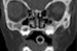
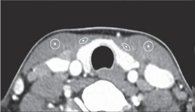
| Compared with unshielded CT image (above), mean attenuations of both superficial neck muscles (strap muscle [squares] and sternocleidomastoid [asterisks]) are higher in shielded image (below), whereas image noises do not show significant difference. Images republished with permission of the American Roentgen Ray Society from AJR 2011; Vol. 196:3, pp. 611-615. |
 |
Lee and his team looked at two groups of patients who were imaged with neck CT but ultimately shown to be without abnormalities. One group had shielding and the other did not. The shields were fabricated from a double layer of bismuth-impregnated latex placed with a 2-cm radiolucent cotton spacer to decrease streak artifacts near the contact surface with the shield. There were no significant demographic differences between the two groups.
For one month in 2007, the researchers applied the shield over the anterior neck of all patients during routine neck CT exams to protect the thyroid gland, they wrote. A month later, the practice was halted and patients once again underwent CT exams without shielding.
The study retrospectively analyzed the results of 84 patients (45 men, 39 women; mean age, 42.3 years; range, 23-71 years), all without abnormalities at CT. Subjects were placed into one of two groups, depending on whether shielding with a cotton spacer was placed over the thyroid before scanning (n = 44) or not (n = 40).
The investigators measured CT attenuation and noise in the strap and sternocleidomastoid (SCM) muscles, as well as superficial radiation dose, using a head CT dose phantom containing ionization chambers located at the 3, 6, 9, and 12 o'clock positions.
An experienced neuroradiologist examined the images using a soft-tissue algorithm on a PACS network (PiView, Infinitt). An ionization chamber and a radiation monitor controller were deployed for dose measurement.
The results showed that CT attenuation rose with use of the commercial bismuth shield.
"These differences between the shielded and unshielded neck CT examinations were statistically significant (p < 0.01), which implies that the use of the shield resulted in increased CT attenuation of the superficial neck muscles compared with the unshielded CT examination," the authors wrote.
Mean CT attenuation and noise values with and without thyroid shielding
|
|||||||||||||||||||||||||||
| Data from Lee et al, AJR 2011, Vol. 196:3, pp. 611-615. | |||||||||||||||||||||||||||
At the same time, noise was unaffected by the use of shielding.
"These differences in noises measured in the superficial neck muscles between shielded and unshielded CT examinations were not statistically significant (p = 0.201 and p = 0.953, respectively)," the study team reported.
The average CT dose index (CTDI) reduction resulting from bismuth thyroid shielding varied by position.
"The phantom study showed that the use of the shield significantly reduced the superficial unshielded dose at the 12 o'clock position compared with the other directions (3, 6, and 9 o'clock positions, p < 0.05)," the authors wrote.
The findings are consistent with previous research showing that shielding substantially reduces radiation dose, particularly in the area just below the shield.
"We noted that the dose-reducing effect by the shield was particularly pronounced at the superficial surface just below the shield (i.e., the 12 o'clock position) compared with the other surfaces, such as lateral or deep to the shield (i.e., the 3, 6, and 9 o'clock positions)," they wrote.
However, the shield also produced increased CT attenuation near the shield during neck CT examinations, while image noise was maintained.
Mean radiation dose by location in 16-cm diameter head dose phantom
|
||||||||||||||||||||
| Data from Lee et al. |
This increased attenuation was also noted in a study by Kalra and colleagues, who found that shielding increased CT attenuation for the shielded surface in an anthropomorphic chest phantom study, which they suggested was likely due to a metal artifact caused by bismuth implanted within the shield.
Metal implants in the CT scan field tend to degrade CT image quality as a result of beam hardening, photon starvation, scattering, and edge gradient effects, Lee and colleagues wrote.
"Our results with neck CT image assessment showed that CT attenuation values were inevitably increased in superficial anterior neck muscles below the shielded surface, although a 2-cm cotton spacer successfully prevented an increase in noise associated with shield application," they added.
In any case, these effects could cause difficulties in the interpretation of neck images, "especially for disease involving the superficial soft-tissue structures adjacent to the thyroid shield, such as cellulitis," they wrote. "Thus, the use of the shield should be carefully considered in CT examinations if the suspected abnormalities are located near the shield."
As for limitations of their research, the investigators noted that they did not scan any pediatric patients, a group that represents the main target of dose-reduction efforts in the thyroid. In addition, radiation exposure was measured only in muscle tissue and not in other structures.
Further investigation is needed to examine the effects of shielding in nonmuscle tissue types, according to the group.
"Both phantom and neck CT image assessment studies have shown that shield application substantially reduces the radiation dose below the shielded surface without an increase in image noise," Lee and colleagues concluded. "However, erroneously increased CT attenuation might be possible. Therefore, care should be exercised for shield application during neck CT examinations, particularly for abnormalities that might be near the shielded surface."