Virtual colonoscopy studies can be read nearly twice as fast using an advanced panoramic view (PV) technique compared to conventional 3D endoscopic views, according to researchers at the Medical University of Vienna. The overall sensitivity for polyp detection is equivalent.
The panoramic technique is one of many reading alternatives for virtual colonoscopy (also known as CT colonography or CTC) that have challenged the standard 3D endoscopic view in recent years. But the generally faster viewing times afforded by these methods have often been offset by reduced polyp sensitivity due to distortion of the mucosa and other drawbacks. The Austrian researchers wanted to see if things might be different with panoramic viewing compared to 3D review and published their findings online before print in the journal European Radiology (October 3, 2010).
"We hypothesized that both interpretation techniques would be equally sensitive in the detection of clinically significant polyps," Thomas Mang, MD, and colleagues wrote. "According to recent trials, primary 2D evaluation is equally as accurate as primary 3D, but potentially less time-consuming. Our study indicates that unidirectional PV interpretation results in a significantly faster 3D CTC evaluation."
When performing a standard primary 3D read, 2D axial views and multiplanar reformats (MPRs) are still necessary to characterize 3D findings and evaluate underdistended or collapsed segments, they wrote. Standard endoscopic 3D views are intended to simulate an optical colonoscopy exam as regards the position of the endoscope, its viewing angle, and intraluminal navigation.
However, optical colonoscopy as such is not a perfect examination because of limited surface visualization, resulting in unseen areas or so-called "blind spots." Therefore, it must be performed twice -- in antegrade and retrograde positions -- to see the lesions hidden behind haustral folds.
Several new 3D projection types have been created to overcome this limitation and increase the surface area visualized, including the PV tool (Siemens Healthcare, Erlangen, Germany). PV is now included in the syngo.via package of software tools.
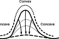
The panoramic technique "renders five faces of a cubic viewing space into one view plane in a continuous fashion," the authors wrote. "Thus, both sides of the haustral folds are displayed in one single fly-through, potentially allowing for unidirectional evaluation which may be more time-efficient."
 |
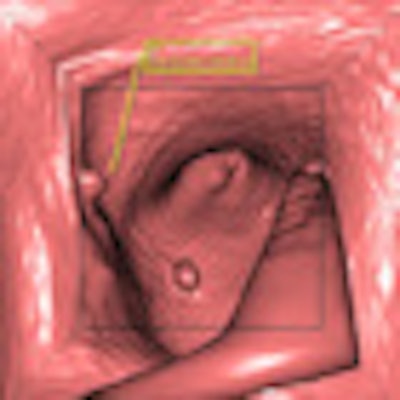
| Colonic mucosa visualized in PV (a component of the syngo.via package). Syngo.via can be used as a standalone device or together with a variety of syngo.via-based software options that are medical devices in their own rights. Images courtesy of Siemens Healthcare. |
 |
The virtual colonoscopy images were acquired prone and supine on a 64-detector-row scanner (Somatom Sensation 64, Siemens) following purgative bowel cleansing and automated CO2 insufflation (ProtoCO2l, Bracco, Milan). Images were acquired at 0.75-mm collimation and 0.5-mm reconstruction increments at 120 kVp and a reference setting of 70 mAs (supine) and 30 mAs (prone) using dose modulation software (Care Dose 4D, Siemens).
Before the initiation of bowel distension, 20 mg of n-butylscopolamine (Buscopan, Boehringer Ingelheim, Ingelheim, Germany) was administered intravenously.
The study examined 150 consecutive CTC patient datasets that were retrospectively reviewed twice by two expert radiologists. The first exam was read using a standard bidirectional view, and the second exam, from six to 15 months later, was read using the unidirectional panoramic view technique, the authors wrote. The results of all exams were validated endoscopically using conventional colonoscopy.
"With regard to polyp detection, we found equivalence with regard to sensitivity of the two techniques for both adenomas and for nonadenomatous polyps," they wrote. "The unidirectional PV interpretation revealed slightly higher overall per-polyp sensitivities compared with bidirectional 3D [standard view (SV)] evaluation (68.2% versus 66.9%)."
Polyp detection sensitivity for SV
|
|||||||||||||||||||||||||||||
Polyp detection sensitivity for PV
|
|||||||||||||||||||||||||||||
Both methods provided equivalent polyp detection, with an equivalence limit set at 5%. However, the panoramic view visualized significantly more of the colonic mucosa (98.9% ± 1.1%; range, 97.0%-99.9%) compared with standard 3D endoluminal viewing (96.2% ± 2.3%; range, 91.4%-98.8%) (p > 0.05).
On the other hand, there were more false positives using the panoramic view: a total of 20 (≤ 5 mm, n = 16; 6-9 mm, n = 4) with bidirectional standard 3D views versus 28 (≤ 5 mm, n = 23; 6-9 mm, n = 5) using the unidirectional retrograde PV.
The mean interpretation time dropped from 14.6 ± 2.5 minutes (range, 9.2-22.8) with standard 3D viewing to 7.5 ± 3.2 minutes (range, 5.0-14.4) using PV (p < 0.0001).
"Our study indicates that unidirectional PV interpretation results in a significantly faster 3D CTC evaluation," the authors wrote. "The mean reading time in our cohort with SV virtual endoscopy for the evaluation of a patient dataset consisting of prone and supine images (mean 14.6 min) was within the range of results published in the literature."
Data evaluation with panoramic viewing in a single direction reduced the mean reading time by almost 50% (7.5 minutes versus 14.6 minutes) compared with a bidirectional SV approach.
"Compared with conventional 3D techniques, the panoramic view displays both sides of haustral folds in a single fly-through for a time-effective single retrograde evaluation," Mang and colleagues wrote.
However, the authors cautioned, even though panoramic view visualizes a large amount of the mucosa in a single fly-through, there may be limitations near sharp turns in the colon -- such as in a tortuous sigmoid colon or at the flexures -- which can lead to lesions being missed.
"Whereas unidirectional SV mainly missed areas behind or between haustral folds, bidirectional SV missed only blind spots between narrow standing haustral folds and in some of the inner areas of colonic flexures," they wrote.
Other potential pitfalls for panoramic viewing include luminal distortion and the short display time for blind spots and the back of the colonic mucosa. A shorter display time could potentially lead to more missed lesions.
The new viewing modality is likely to have a long learning curve; however, over time perception is likely to improve. More studies are needed to evaluate the feasibility of this and similar tools, they wrote.
The detected prevalence of adenomas 6 mm and larger and advanced lesions 10 mm and larger was in good agreement with lesions in the symptomatic German population, a prevalence documented in more than 2 million studies.
"We conclude that 3D unidirectional retrograde data evaluation with a 3D panoramic view significantly reduces the reading time with no significant differences in sensitivity compared with bidirectional 3D standard view," Mang and colleagues wrote. "This technique visualizes the complete colonic mucosa in a unidirectional fly-through, whereas standard 3D tools require bidirectional fly-through to achieve complete visualization."
By Eric Barnes
AuntMinnie.com staff writer
February 11, 2011