Although virtual colonoscopy practice has always focused on adenoma detection and colon cancer prevention, the exam is proving to be formidable in the staging of diverticular disease, a scourge of fast-food Western nations that affects most of their citizens by age 60.
Virtual colonoscopy (also known as CT colonography or CTC) has several advantages in this setting. Unlike conventional colonoscopy, VC is unaffected by tortuous colon morphology that can be associated with diverticular disease. Diverticular disease is also well characterized by MR and, in some cases, ultrasound, but MDCT remains the gold standard.
Diverticular disease affects a large number of individuals who are also VC's prime screening target, according to Dr. Patrick Rogalla, a professor of radiology from Charité University Hospital in Berlin. He spoke at Stanford University's International Symposium on Multidetector-Row CT in May.
A lifestyle disease
"Diverticular disease has a strong association with the Western lifestyle, and also age," Rogalla said. "It has an inverse correlation with the fiber content in the diet and a positive correlation with the amount of red meat in the diet."
The effects are compounded to the point where people whose diets are both low in fiber and high in red meat consumption have three times the risk of developing diverticulosis as the general population, he said.
Age also is a factor: The incidence of diverticular disease rises "from less than 10% of young individuals to more than 60% in elderly patients, and that is certainly of great importance to our society," Rogalla said.
Far from a mere annoyance, more than one-third of diverticulosis cases will eventually progress to diverticulitis, a potentially dangerous condition that affects about 10 in every 100,000 individuals and is responsible for upward of 200,000 hospital admissions annually in the U.S.
"As you can imagine, this is a big healthcare problem," he said. "Approximately 25% of patients with acute diverticulitis will develop life-threatening complications such as hemorrhage or perforation."
The clinical presentation of acute diverticular disease is typified by the triad of lower abdominal pain, fever, and leukocytosis, Rogalla said. Two staging systems in common use see manifestations a bit differently. Peritonitis may or may not be present in generalized local disease, he said. Sometimes a palpable roll is present in the abdomen, along with tenesmus, dysuria, or pneumaturia.
 |
| All images courtesy of Dr. Patrick Rogalla. |
Generally, diverticular disease is confined to the sigmoid colon, but it is a right-sided disease in 15% of all cases, Rogalla said. In both presentations, there is a trend toward conservative management of acute disease.
Imaging for diverticular disease
"Barium enema used to be the standard of reference, but with the advent of CTC we don't even offer that study anymore in our institution," Rogalla said.
Among the latest available comparison studies, Ambrosetti and colleagues in 2000 compared iodine contrast enema to CT in a prospective study of 420 patients with complicated diverticulitis, all with surgical confirmation. Sensitivity for disease detection was 92% for barium enema versus 98% for CT (p < 0.01). And while barium enema found 29% of the abscesses, CT detected them with 100% sensitivity (Diseases of the Colon and Rectum, October 2000, Vol. 43:10, pp. 1363-1367).
CT is the exam of choice for diverticulitis evaluation, not only for its superior sensitivity, but for its ability to detect severe infection, "especially when an abscess is associated with the disease," the authors concluded.
Ultrasound is a good modality for detecting the complications of diverticular disease, and MR images are quite similar to those of CT, Rogalla said. CT also provides images very similar to those of optical colonoscopy, but noninvasively.
CT is considered the modality of choice for detecting the complications of diverticular disease, including free air, perforation, fistula, abscess formation, peritonitis, and hemorrhage. In a 2008 study, Heverhagen and colleagues found the following features in their cohort of patients with advanced disease:
- Pericolic fatty stranding (98%)
- Diverticula (84%)
- Wall thickening (70%)
- Abscess (47%)
- "Lucky hat" sign (30%)
- Free air (16%)
- Fistula (14%)
- Obstruction (12%)
- Ureteral obstruction (7%)
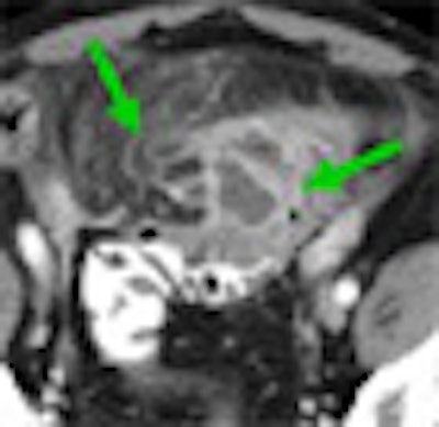
In cases with local complications such as hemorrhage and perforation, both CT and MRI often demonstrate the "lucky hat" sign, which indicates the active migration of omental fat, which drives local inflammation, Rogalla said.
 |
| Locally complicated cases with hemorrhage and perforation often reveal a "lucky hat" sign, indicating active migration of omental fat that drives local inflammation. Extracolonic abscess formation indicates stage IIB disease by the Hanson/Stock staging method and stage II by the modified Hinchey system. |
Importantly, "CT has the highest sensitivity for tiny air bubbles, which would not be detected on a conventional x-ray, ultrasound, or MRI," he said. "That's why CT is so important in this disease to detect the complications."
 |
| CT's most important advantage in imaging diverticular disease is its high sensitivity for air bubbles indicating colonic perforation. At left, bubble at CT reveals a single perforation in the colonic mucosa, confirmed in postsurgery specimen (right). |
Two staging systems
Two staging methods are in widespread use: the Hanson/Stock staging method popular in Europe and the modified Hinchey (Wasvary) technique dominant in the U.S., Rogalla said.
 |
| Two staging methods are in widespread use: the Hanson/Stock staging method popular in Europe and the modified Hinchey dominant in the U.S. All images courtesy of Dr. Patrick Rogalla. |
Per the Hinchey method, for example, complicated disease is classified as stage I as long as the complication is local, he said. Once an abscess is found at CT, patients are potential candidates for CT-guided catheter placement under fluoroscopy to drain the abscess.
 |
| Diverticular disease above reveals wall thickening and stranding in the pericolonic fatty tissue at CT. Case below shows moderate obstruction and acute inflammation. Both are classified as stage 0 by the Hanson/Stock method and as stage I by the modified Hinchey method. |
 |
"Once we have distant metastatic [infection] sites, it will be stage II according to the Hinchey classification, although the local findings are minimal," Rogalla said. "When we see abscess formation, sometimes the sigmoid colon looks almost normal, but in almost all cases that's the source of the infection." Once again the abscesses can be drained at CT-guided fluoroscopy, he said.
Diverticulitis can sometimes form fistulas in neighboring organs, mimicking a mass in the urinary bladder, according to Rogalla.
It's important to be aware of differential diagnoses including ascites, colorectal cancer, and colitis, all of which can mimic diverticular disease, he said. Appendagitis needs to be ruled out before surgery because it resolves itself.
Some surgeons choose to operate a little earlier for right-sided disease, for which hernia should be ruled out, Rogalla said. In cases where diverticular disease extends to the small bowel, it, too, may need to be resected.
Conservative treatment versus surgery
"With respect to therapy, there is a great degree of uncertainty, and the pendulum goes back and forth," Rogalla said. "Sometimes the surgeons want to operate very early; some are on the conservative side. But there is some agreement across the Atlantic that early stages of diverticular disease with pericolic fat stranding should undergo conservative treatment, with no surgical option. Once you see massive peritonitis, as well as free air, the patient should undergo emergency surgery."
The treatment decision is unclear for less severe cases, including Hinchey stages II and IB with local and distant abscess formation, he said. Sometimes a case with a large intramural abscess can be treated with antibiotics, rendering the surgery easier.
"Some surgeons would go right away toward a two-stage surgical procedure to take the sigmoid out, because we do see local intramural abscess formation and a lot of infiltration regarding abscess formation with the lucky hat sign," he said.
Taking a conservative approach in one recent case of stage II disease, Rogalla's institution chose an eight-day antibiotic regimen, which returned the sigmoid to almost normal appearance at VC. As a result of antibiotic therapy, he said, the patient was able to undergo single-stage elective surgery rather than the emergency two-stage surgery that would have been required for immediate surgery -- with important advantages for the patient.
 |
| Acute cases sometimes benefit from conservative management before surgery. Diverticular disease with intramural abscess at CT before (above) and after (below) antibiotic treatment, followed by single-stage elective resection of the sigmoid colon. |
 |
MDCT is the method of choice for staging diverticular disease, though there are alternatives, because each of the other modalities presents disadvantages, Rogalla concluded. Radiologists should understand both the European and U.S. staging criteria, he said.
"In general, there is a trend toward conservative therapy, with CT-guided drainage and downstaging with antibiotics -- then elective single-stage surgery and primary anastomosis -- because patients benefit much more from this kind of conservative approach," Rogalla said.
By Eric Barnes
AuntMinnie.com staff writer
September 22, 2009
Related Reading
MR colonography separates benign from malignant colonic strictures, January 16, 2009
Unusual VC regimen a success in the elderly, November 27, 2007
MDCT finds cause of acute abdomen more often with MPR, November 29, 2006
Rare venous malformations look like polyps on VC, April 12, 2006
Diverticular disease doesn't impede VC in Wisconsin study, November 28, 2005
Copyright © 2009 AuntMinnie.com