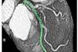
Computer-aided detection (CAD) developer R2 Technology has launched a pulmonary artery patency exam (PE) tool.
Designed to help physicians detect potential pulmonary artery obstructions or filling defects such as emboli or tumors during multislice CT exams, the tool examines patency following a multislice CT pulmonary angiogram, according to the Sunnyvale, CA-based vendor. It then performs an anatomical segmentation process, providing automatic calculations of vessel diameter, percent occlusion, and size of the detected obstruction, R2 said.
The tool is available with version 2.0 of R2's CT Lung CAD system.
By AuntMinnie.com staff writers
June 16, 2005
Related Reading
R2 updates mammo CAD, June 3, 2005
R2 debuts ImageChecker 2.0 for CT, May 17, 2005
R2 adds to executive lineup, May 13, 2005
iCAD, R2 in patent dispute, April 28, 2005
Vital Images, R2 form alliance, April 26, 2005
Copyright © 2005 AuntMinnie.com


![Images show the pectoralis muscles of a healthy male individual who never smoked (age, 66 years; height, 178 cm; body mass index [BMI, calculated as weight in kilograms divided by height in meters squared], 28.4; number of cigarette pack-years, 0; forced expiratory volume in 1 second [FEV1], 97.6% predicted; FEV1: forced vital capacity [FVC] ratio, 0.71; pectoralis muscle area [PMA], 59.4 cm2; pectoralis muscle volume [PMV], 764 cm3) and a male individual with a smoking history and chronic obstructive pulmonary disorder (COPD) (age, 66 years; height, 178 cm; BMI, 27.5; number of cigarette pack-years, 43.2, FEV1, 48% predicted; FEV1:FVC, 0.56; PMA, 35 cm2; PMV, 480.8 cm3) from the Canadian Cohort Obstructive Lung Disease (i.e., CanCOLD) study. The CT image is shown in the axial plane. The PMV is automatically extracted using the developed deep learning model and overlayed onto the lungs for visual clarity.](https://img.auntminnie.com/mindful/smg/workspaces/default/uploads/2026/03/genkin.25LqljVF0y.jpg?auto=format%2Ccompress&crop=focalpoint&fit=crop&h=112&q=70&w=112)


