SALT LAKE CITY - Many Winter Olympic sports involve breathtaking, high-flying maneuvers such as freestyle aerial skiing. It was one of these moves, unfortunately, that resulted in a midfoot fracture-dislocation, including a navicular fracture and dislocation between the lateral cuneiform and the cuboid.
The images below were acquired on an Mx8000 quad-detector CT scanner (Philips Medical Systems, Bothell, WA) at the University of Utah hospital, located a few minutes away from the Polyclinic. The images were reformatted on a Marconi PACS station by the radiologist. Reformatting enables the radiologist to choose the optimal plane and angulation.
 |
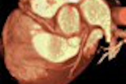
| Direct coronal CT image through the midfoot, demonstrating dislocation between the lateral cuneiform and the cuboid. Soft-tissue algorithms are routinely used in addition to bone algorithm in CT trauma evaluation to enable evaluation of tendons and ligaments. This soft-tissue image shows tiny avulsion fragments dorsally due to avulsion of the interosseous ligaments between the cuneiform and cuboid. |
 |
| Sagittal reformatted image through the lateral aspect of the foot shows inferior subluxation of the cuboid (arrow) relative to the fourth metatarsal. |
 |
| Sagittal reformatted image through the medial aspect of the foot demonstrates the fracture (arrow) of the inferior portion of the navicular. |
By Dr. Julia Crim
AuntMinnie.com contributing writer
February 22, 2002
Dr. Crim is chief of musculoskeletal imaging at the Olympic Polyclinic.
Related Reading
Scenes from the Polyclinic: syndesmosis, February 20, 2002
Scenes from the Polyclinic: A case study in the ACL-deficient knee, February 19, 2002
Medical imaging goes for gold at Olympic Polyclinic, February 8, 2002
Copyright © 2002 AuntMinnie.com