
Portable chest x-ray images appear comparable to those acquired with standard digital systems in patients with tuberculosis (TB) -- and this could be good news for TB screening efforts, according to a group in India.
Chest x-ray is an important screening tool for pulmonary TB, yet accessibility to it is a challenge in difficult-to-reach and underserved populations. This can potentially be overcome by deploying portable digital x-ray machines, the authors suggest.
"However, these portable x-ray machines need to be validated before their deployment in the field," wrote first author Dr. Raj Kamal of the National Jalma Institute for Leprosy in Agra, and colleagues. The study was published April 15 in the Indian Journal of Medical Research.
To that end, the group tested image quality as evaluated by two radiologists between a model called Mine 2 (LabIndia Healthcare), a portable handheld x-ray system weighing 1.8 kg, and digital x-ray units routinely used in their health facilities.
The researchers recruited 100 participants with suspected pulmonary TB from outpatient clinics and a community health center in Agra. Each participant underwent two chest x-rays, once with each type of system. Both sets of deidentified images were then independently read by two radiologists blinded to the type of machine used. The primary outcome was agreement between the quality of images produced by the two systems.
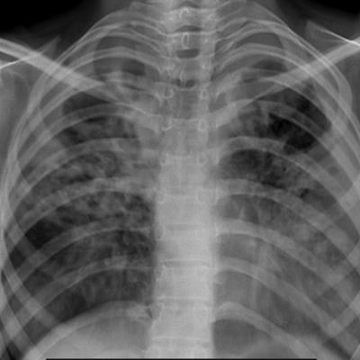
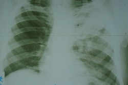
 Chest x-ray images for the same individual with evidence of tuberculosis using (A) the Mine 2 machine, and (B) a standard digital x-ray machine. Image courtesy of the Indian Journal of Medical Research through CC BY 4.0.
According to the findings, the intraobserver (radiologist) agreements regarding the status of 15 chest x-ray parameters between the images ranged between 74% and 100%, with an unweighted mean of 87.2%, the group reported. In addition, in an overall comparison of image quality (on a scale of one to 10, with 10 denoting highest quality), the handheld x-ray images had a score of 9 and the digital chest x-ray images had a score of 8, the researchers stated.
"The current study shows that a handheld x-ray machine, which is easy to use and can potentially be carried to any area, produces x-ray images with quality that is comparable to digital x-ray machines routinely used," the group wrote.
While promising, experiences of using handheld x-ray machines for dental purposes have shown that certain issues arise in their use, such as operator movements, radiation protection of the operator, operator fatigue, and disinfection, they noted.
"Further research is needed to evaluate the efficacy and effectiveness of handheld chest x-devices, as well as to examine implementation issues such as feasibility, adoption, and cost-effectiveness," they wrote.


![A normal mammogram confirmed by three-year radiologic follow-up illustrates reader-marked regions of interest (ROIs) during (A) unaided (round 1) and (B) artificial intelligence (AI)–assisted (round 2) reading. Each colored dot represents an ROI for recall by a human reader. Readers could mark more than one ROI per case, represented by multiple dots of the same color. During AI-assisted reading, the AI system displayed three visible prompts: two with suspicion of malignancy scores of 35% (left mediolateral oblique [L MLO] and craniocaudal [L CC]) and one with a suspicion of malignancy score of 10% (right craniocaudal [R CC]), shown as polygonal overlays. Without AI, six of 10 readers (60%) marked a false-positive ROI. With AI assistance, this fell to two of 10 (20%). R MLO = right mediolateral oblique.](https://img.auntminnie.com/mindful/smg/workspaces/default/uploads/2026/07/2026-07-14-radiology-mammogram-ai-auto-bias.H0bYO8QlWs.jpg?auto=format%2Ccompress&dpr=2&fit=crop&h=167&q=70&w=250)


