
Estimating bone age in children using dual-energy x-ray absorptiometry (DEXA) is unreliable, says a group of researchers in Mexico. The team compared the approach to standard x-ray imaging in a study published August 17 in the Journal of Clinical Densitometry.
Epidemiological researchers compared bone age estimates using DEXA and x-ray exams in a large number of children between the ages of 5 and 16 who underwent hand imaging at the Hospital Infantil de Mexico in Mexico City. They found a high agreement among readers using both techniques but that bone age estimates varied among age groups.
"We identified that in [DEXA] images, bone age tends to be overestimated," wrote corresponding author Dr. Patricia Clark, PhD, a rheumatologist and head of the hospital's clinical epidemiology research unit, and colleagues.
Bone age is a measure of developmental age, or physiological maturity, which is a more accurate representation than chronological age. It is particularly helpful in the clinical workup of children with growth or puberty disorders, for instance.
Standard bone age estimates to evaluate bone age in pediatric patients are qualitative and based on the doctor's recognition of maturation features on hand x-rays compared with a reference atlas. As an alternative, some researchers have suggested that DEXA imaging may be a valuable alternative, especially since it allows for a reduction in radiation dose in vulnerable pediatric patients, according to the authors.
Yet so far, studies comparing the approaches have been small, they wrote. Thus, in this study, Clark and colleagues explored the use of the two approaches in a large sample size stratified by age and sought to determine if DEXA might replace x-ray imaging in clinical practice.
The researchers culled images of 711 children and adolescents (307 female) aged 5 to 16 who underwent evaluations of the hand in two previous clinical trials. For all subjects, hand and wrist images for both techniques were obtained during the same day. For every hand and wrist reading, both x-ray and DEXA images were performed by two pediatric endocrinologists.
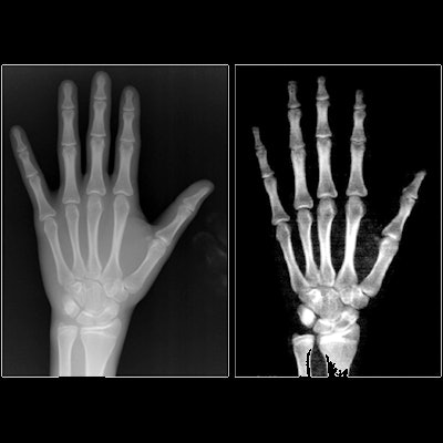
 An example of an x-ray image (left) and DEXA image (right) of nondominant hand used in the study. The images are from a male patient (chronological age: 15.4 years; bone age by x-ray, 15.5 years). Image courtesy of the Journal of Clinical Densitometry.
The researchers found a high concordance between bone age estimations of both imaging techniques in the total sample, with agreement between readers greater than 0.98. The mean difference in bone age estimates between both techniques was 0.34 years, with agreement limits contained within ± 2 years.
Yet on Bland and Altman plots (displays of the relationship between two paired variables), the researchers observed overestimations of bone age measurements in DEXA, they wrote. Bone age estimate differences were less than six months in groups of 5- to 8-year-old children as well as groups of 14- to 16-year-old adolescents, but the estimate differences increased to 0.9 years in ages 9 to 13.
"With this information, the implementation of bone age measurement using [DEXA] images appears not to be reliable," the authors wrote.
The authors hypothesized that the lower beam energy of DEXA implies a lower quality image compared to conventional x-ray imaging, and that this factor translated into a high probability of bone age overestimation in DEXA images in their study compared with x-rays.
Nonetheless, DEXA imaging remains a promising alternative, especially given its promise of lower patient radiation exposure, they wrote.
"From our results we may say that, if the clinical purpose of the bone age evaluation requires a precision lower than 0.5 years, the classical bone age evaluation technique using conventional X-ray imaging must be suggested," Clark and colleagues concluded.


![A normal mammogram confirmed by three-year radiologic follow-up illustrates reader-marked regions of interest (ROIs) during (A) unaided (round 1) and (B) artificial intelligence (AI)–assisted (round 2) reading. Each colored dot represents an ROI for recall by a human reader. Readers could mark more than one ROI per case, represented by multiple dots of the same color. During AI-assisted reading, the AI system displayed three visible prompts: two with suspicion of malignancy scores of 35% (left mediolateral oblique [L MLO] and craniocaudal [L CC]) and one with a suspicion of malignancy score of 10% (right craniocaudal [R CC]), shown as polygonal overlays. Without AI, six of 10 readers (60%) marked a false-positive ROI. With AI assistance, this fell to two of 10 (20%). R MLO = right mediolateral oblique.](https://img.auntminnie.com/mindful/smg/workspaces/default/uploads/2026/07/2026-07-14-radiology-mammogram-ai-auto-bias.H0bYO8QlWs.jpg?auto=format%2Ccompress&dpr=2&fit=crop&h=167&q=70&w=250)


