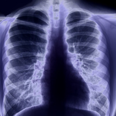
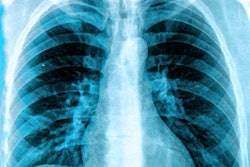
A guideline detailing the use of chest x-ray for patients suspected of having the COVID-19 respiratory disease has been published by a group from the University of California, Los Angeles.
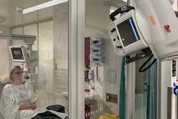
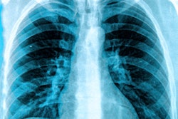
CT and, to a lesser extent, ultrasound have been the imaging tools used most often for COVID-19 diagnosis. But x-ray is also seen as having value due to its ease of use and ability to be moved to the patient's bedside.
The thoracic and acute care imaging sections at UCLA's department of radiology developed the UCLA Chest X-Ray COVID-19 Guideline to help physicians and radiologists across a variety of clinical settings identify which radiographic findings are most likely due to COVID-19 involvement. The guidelines also cover which other clinical conditions should be considered in these cases.
The guidelines are available on the UCLA department of radiology's website.






![A normal mammogram confirmed by three-year radiologic follow-up illustrates reader-marked regions of interest (ROIs) during (A) unaided (round 1) and (B) artificial intelligence (AI)–assisted (round 2) reading. Each colored dot represents an ROI for recall by a human reader. Readers could mark more than one ROI per case, represented by multiple dots of the same color. During AI-assisted reading, the AI system displayed three visible prompts: two with suspicion of malignancy scores of 35% (left mediolateral oblique [L MLO] and craniocaudal [L CC]) and one with a suspicion of malignancy score of 10% (right craniocaudal [R CC]), shown as polygonal overlays. Without AI, six of 10 readers (60%) marked a false-positive ROI. With AI assistance, this fell to two of 10 (20%). R MLO = right mediolateral oblique.](https://img.auntminnie.com/mindful/smg/workspaces/default/uploads/2026/07/2026-07-14-radiology-mammogram-ai-auto-bias.H0bYO8QlWs.jpg?auto=format%2Ccompress&fit=crop&h=112&q=70&w=112)


