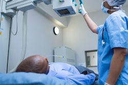
Taking patient photographs at the point of care to attach them to portable radiographs is technically feasible and also confers numerous advantages for patient care, according to a study published in the January edition of American Journal of Roentgenology.
A team of researchers led by Dr. Srini Tridandapani, PhD, from Emory University in Atlanta mounted wide-angle smart cameras (Wide Angle FOV160°, SainSmart) on portable radiography machines (DRX Revolution, Carestream Health) and then tested the technology at Emory, a large academic medical center.
They hoped to build on previous research that found that attaching patient photos to x-rays conferred a variety of benefits, such as fewer wrong-patient error and potentially other advantages like increased empathy for patients thanks to the personalization of their radiographs (AJR, January 2020, Vol. 214:1, pp. 68-71).
The original goal of the project was to reduce wrong-patient errors, and Tridandapani and colleagues noted that previous research led the team to expect that a wrong-patient error would occur once in every 10,000 studies. In the current study, however, investigators noticed one error within the first 300 cases obtained using the photo identifiers and two errors overall in the first 8,000 cases.
One advantage of capturing digital photographs at the time of radiography examinations is that the photographs offer confirmation that technologists are employing proper placement/positioning techniques and carrying out appropriate precautions to shield patients.
One possible disadvantage is that the use of photographs may increase interpretation time, the authors noted. Investigators reported, however, that they found the use of the photographs accelerated interpretation in the case of chest and abdominal portable radiography. This is because the photographs displayed the external portions of lines and tubes, avoiding possible confusion in separating internal from external portions when radiographs alone are used.
When it comes to extremity radiographs obtained in the cases of trauma or to assess for osteomyelitis in patients with diabetic foot ulcers, photographs can assist radiologists by allowing them to focus their attention on the portion of the bone on the radiograph underlying the visible soft-tissue injury or ulcer.
The laterality information offered in the photographs also is of value to musculoskeletal radiologists who can sometimes be uncertain about whether a right or left extremity has been imaged. A photograph can display patient orientation with the radiographic plate behind the limb being radiographed, confirming that the correct side is being imaged.
The authors noted that patient privacy and security are paramount concerns with the system, and they have taken measures such as deleting raw photographs immediately from the cameras once retrieved by the integration server over the hospital's secure wireless network. Moreover, each camera is registered by the hospital IT department, so that it can only communicate with and be accessed by the integration server.
Technologists had expressed concerns about the use of wide-angle cameras compromising their privacy, but education about the objective of the technology allayed these concerns for the most part.
One of the requirements to ensure successful interpretation is having even lighting in a room, with the authors noting that dim and uneven room lighting can produce photographs that are not evenly lit, making interpretation challenging.


