DUBAI - New details have emerged about the Arab School of Radiology (ASOR). The official launch is now scheduled for the 7th Pan Arab Radiology Congress, to be held in Doha, Qatar, on 17-21 April, and in September ASOR will stage a three-day workshop in Abu Dhabi, United Arab Emirates, on breast imaging.
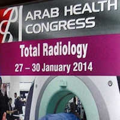
ASOR is being promoted at this week's Arab Health congress by Lebanese-based staff from Medical Professionals. In July 2013, the founder of ASOR, the Pan Arab Association of Radiological Societies (PAARS), appointed the company to launch and administer the new organization.
"This is a serious step forward for this broad medical community, which has historically not had any practical venue for advancement, in comparison to peers in Western Europe, North America, and elsewhere, whom have long benefitted from dedicated bodies such as the RSNA, ESOR (European School of Radiology), and the SFR (French Society of Radiology) to name a few," said Dr. Maurice Haddad, executive officer of ASOR and secretary general of PAARS.
ASOR will be dedicated to the advancement and continuing education of radiologists and radiographers in the Middle East and North Africa with lectures, workshops, and other training solutions.The plan is for the group to become a super-regional body providing scientific and practical content to the region's radiology professionals, taking account of their varied levels of skills and aptitude. Its website is currently under construction and will also be unveiled in April.
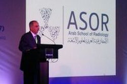
 This distinctive new logo has been developed for the Arab School of Radiology.
The course director for ASOR's Abu Dhabi workshop will be Dr. Nathalie Duchesne, a breast radiologist at Hôpital du Saint-Sacrement in Quebec City, Canada. The registration fee is $730 U.S. (534 euros) for a radiologist and $450 U.S. (329 euros) for a radiographer.
"We have historically been disappointed by the scientific content evident in most of the region's medical congresses and exhibitions, and have conceived the ASOR jointly between all of the heads of the various Arab radiology societies as a well overdue necessity, and as a complement to the annual Arab Radiology Congress," said Dr. Assaad Mohanna, former president of the Lebanese Society of Radiology and assistant secretary general of PAARS.
Haddad, who is based in the department of radiology at the American University of Beirut Medical Center,was the lead author of an influential position paper about the status of radiology in the Arab world (The Lebanese Medical Journal, J Med Liban, 2007 April-June; Vol. 55:2, pp. 94-98). He reported that the first x-ray apparatus was installed in 1898 in Beirut at the Prussian Hospital, which changed its name to the American University of Beirut in 1920. The first chest radiographs were produced after a long exposure time in 1900 by Reverend Père Maurice Collangettes at the Faculté Française de Médecine in Beirut and by Dr. Arthur Bacon and Brahim Youssef Succar, the first radiographer at the x-ray laboratory of the Prussian Hospital.
"The input and support of eminent immigrant Arab professors working in European and North American universities is of paramount importance in maintaining academic activities, particularly in these difficult times when scholars from the Western hemisphere are discouraged from traveling to the Middle East for security reasons," Haddad noted.
He stresses that even though the Arab world is rich in natural and human resources, it has suffered for a long time from many devastating wars and sanctions that have hindered development and frustrated its people, including radiologists. Major problems and challenges in health development still need to be addressed through better planning and healthcare management, and for radiology to move forward, increased inter-Arab cooperation is essential, he said.